Millipore Sigma Vibrant Logo
Attention: We have moved. Merck Millipore products are no longer available for purchase on MerckMillipore.com.Learn More

MABF2075-100UL Anti-MR1 Antibody, clone 26.5

MABF2075-100UL
100 µL  
Purchase on Sigma-Aldrich

Přehled

Replacement Information
Description
Catalogue NumberMABF2075-100UL
DescriptionAnti-MR1 Antibody, clone 26.5
Alternate Names
  • Major histocompatibility complex class I-related gene protein
  • MHC class I-related gene protein
  • Class I histocompatibility antigen-like protein
Background InformationMajor histocompatibility complex class I-related gene protein (UniProt: Q95460; also known as MHC class I-related gene protein, Class I histocompatibility antigen-like protein, MR1) is encoded by the MR1 gene (Gene ID: 3140) in human. MR1 is a single-pass membrane protein that is ubiquitous in its distribution and can heterodimerize with B2M. It serves as an antigen-presenting molecule specialized in presenting microbial vitamin B metabolites. It is involved in the development and expansion of a small population of T-cells expressing an invariant T-cell receptor alpha chain known as mucosal-associated invariant T-cells (MAIT). MAIT lymphocytes are preferentially located in the gut lamina propria and may be involved in monitoring commensal flora or serve as a distress signal. MR1-restricted T cells can discriminate between microbes in a TCR-dependent manner. However, these cells are shown to be severely reduced in circulation in subjects with chronic HIV-1 infection. MR1 is synthesized with a signal peptide (aa 1-22), which is subsequently cleaved off in the mature form. The mature form has an extracellular domain (aa 23-302), a transmembrane domain (aa-303-323), and a short cytoplasmic tail (aa 324-341). Its ligand binding region is localized to amino acids 23-201. Five different isoforms of MR1 have been described that are produced by alternative splicing. (Ref.: Young, MH et al., (2013). PLOS one 8(1); e53789; Leeansyah, E., et al. (2013). Blood. 121(7); 1124-1135).
References
Product Information
FormatPurified
PresentationPurified mouse monoclonal antibody IgG2a in PBS without azide.
Applications
ApplicationAnti-MR1, clone 26.5, Cat. No. MABF2075, is a mouse monoclonal antibody that detects MHC class I-related gene protein and has been tested for use in Flow Cytometry, Function Analysis, and Immunoprecipitation.
Key Applications
  • Flow Cytometry
  • Function Assay
  • Immunoprecipitation
Application NotesImmunoprecipitation Analysis: A representative lot immunoprecipitated MR1 in Immunoprecipitation applications (Huang, S., et. al. (2005). J Biol Chem. 280(22):21183-93).

Flow Cytometry Analysis: A representative lot detected MR1 in flow cytometry applications (Huang, S., et. al. (2005). J Biol Chem. 280(22):21183-93; Gold, M.C., et. al. (2010). PLoS Biol. 8(6):e1000407; Huang, S., et. al. (2009). Proc Natl Acad Sci U S A. 106(20):8290-5).


Inhibits Activity/Function Analysis: A representative lot inhibited the activation of mucosal-associated invariant T (MAIT) cells by MR1 protein. (Huang, S., et. al. (2009). Proc Natl Acad Sci U S A. 106(20):8290-5).
Biological Information
ImmunogenGST-tagged recombinant fragment corresponding to 280 amino acids from the extracellular domain of human MHC class I-related gene protein.
Epitopeextracellular domain
Clone26.5
ConcentrationPlease refer to lot specific datasheet.
HostMouse
SpecificityClone 26.5 specifically detects Major histocompatibility complex class I-related gene protein (MR1). It targets an epitope with in the extracellular domain.
IsotypeIgG2aκ
Species Reactivity
  • Rat
  • Human
  • Mouse
  • Bovine
Species Reactivity NoteHuman, Bovine, Mouse, Rat.
Antibody TypeMonoclonal Antibody
Entrez Gene Number
Gene Symbol
  • MR1
Purification MethodProtein G purified
UniProt Number
Molecular Weight39.37 kDa calculated.
Physicochemical Information
Dimensions
Materials Information
Toxicological Information
Safety Information according to GHS
Safety Information
Product Usage Statements
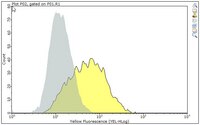
Quality AssuranceEvaluated by Flow Cytometry in MR1-transfected WT3 mouse embryonic fibroblasts.

Flow Cytometry Analysis: 1 µg of this antibody detected MR1 in one million MR1-transfected WT3 mouse embryonic fibroblasts.
Usage Statement
  • Unless otherwise stated in our catalog or other company documentation accompanying the product(s), our products are intended for research use only and are not to be used for any other purpose, which includes but is not limited to, unauthorized commercial uses, in vitro diagnostic uses, ex vivo or in vivo therapeutic uses or any type of consumption or application to humans or animals.
Storage and Shipping Information
Storage ConditionsStable for 1 year at -20°C from date of receipt. Handling Recommendations: Upon receipt and prior to removing the cap, centrifuge the vial and gently mix the solution. Aliquot into microcentrifuge tubes and store at -20°C. Avoid repeated freeze/thaw cycles, which may damage IgG and affect product performance.
Packaging Information
Material Size100 µL
Transport Information
Supplemental Information
Specifications
Global Trade Item Number
Katalogové číslo GTIN
MABF2075-100UL 04054839638602