Millipore Sigma Vibrant Logo
Attention: We have moved. Merck Millipore products are no longer available for purchase on MerckMillipore.com.Learn More

MABN1835 Anti-DYRK1A Antibody, clone 7F3

MABN1835
100 μg  
Purchase on Sigma-Aldrich

Speciální nabídky

Přehled

Replacement Information

Speciální nabídky

Tabulka spec. kláve

Species ReactivityKey ApplicationsHostFormatAntibody Type
H, Mk, RIH(P), WB, IF, ICCMPurifiedMonoclonal Antibody
Description
Catalogue NumberMABN1835
DescriptionAnti-DYRK1A Antibody, clone 7F3
Alternate Names
  • Dual specificity tyrosine-phosphorylation-regulated kinase 1A
Background InformationDual specificity tyrosine-phosphorylation-regulated kinase 1A (EC 2.7.12.2; UniProt Q63470; also known as Dual specificity YAK1-related kinase, MNBH, Protein kinase minibrain homolog, RP86) is encoded by the Dyrk1a (also known as Dyrk) gene (Gene ID 25255) in rat species. DYRK1A belongs to a family of conserved dual-specificity tyrosine-phosphorylated and regulated kinases (DYRKs) within the CMGC (CDK, MAPK, GSK, and CLK) group of eukaryote kinome. DYRK family members share a conserved kinase domain and an adjacent DYRK-homology domain or DH-box (DDDNXDY), wihile differing in their N- and C-terminal regions. DYRK1A is regulated by autophosphorylation on Ser, Thr and Tyr residues, but catalyzes phosphorylation of a broad-range of substrates only on Ser/Thr residues within the RPX(S/T)P motif with a preference for Pro at the +1 position (X). Known DYRK1A substrates include transcription factors (e.g. FKHR, CREB, Gli1, NFAT), splicing factors (e.g. cycling L2, SF3b/SAP155), glycogen synthase, as well as multiple proteins engaged in endocytosis in neurons, including dynamin and synaptojanin 1. DYRK1A is also implicated in promoting the formation of neurotoxic Aβ peptides by phosphorylating APP and presenilin-1. Human DYRK1A gene is located in the Down syndrome critical region of chromosome 21 and the protein plays an essential role in the development of the central nervous system. DYRK1A haploinsufficiency and mutations are the causes of intellectual disability (ID), microcephaly and dysmorphic features in human, while transgenic mice carrying extra copies of the gene exhibit learning defects and motor abnormalities.
References
Product Information
FormatPurified
PresentationPurified mouse IgG2bκ in buffer containing 0.1 M Tris-Glycine (pH 7.4), 150 mM NaCl with 0.05% sodium azide.
Quality LevelMQ100
Applications
Application
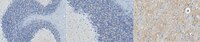
Anti-DYRK1A, clone 7F3, Cat. No. MABN1835, is a highly specific mouse monoclonal antibody that targets DYRK1A and has been tested in Immunocytochemistry, Immunofluorescence, Immunohistochemistry, and Western Blotting.
Key Applications
  • Immunohistochemistry (Paraffin)
  • Western Blotting
  • Immunofluorescence
  • Immunocytochemistry
Application NotesImmunohistochemistry Analysis: A 1:50 dilution from a representative lot detected DYRK1A in rat thalamus tissue section.

Immunocytochemistry Analysis: A representative lot, pre-conjugated with Alexa Fluor® 568, immunostained the soma and processes of 2% formaldehyde-fixed, 0.05% saponin-permeabilized E-18 rat hippocampal neurons by fluorescent immunocytochemistry. DYRK1A immunoreactivity was more abundant in nuclei than in the cytoplasm. Co-localization of DYRK1A and clathrin heavy chain (CHC) immunoreactivity was very prominent in the processes and less significant in the soma (Murakami, N., et al. (2012). PLoS One. 7(4):e34845).

Immunofluorescence Analysis: A representative lot detected DYRK1A cellular localization in GFAP-positive astrocytes and in vimentin-positive endothelial cells by dual fluorescent immunohistochemistry staining of 10% formalin-fixed, PEG 1000-embeded human brain tissue sections (Wegiel, J., et al. (2004). Brain Res. 1010(1-2):69-80).

Immunohistochemistry Analysis: A representative lot detected DYRK1A immunoreactivity in paraffin-embedded fetal (20-week gestation) Down syndrome (DS) white matter associated with structures resembling the omnipresent cytoskeleton filamentous network in axons (Dowjat, K., et al. (2012). J. Neuropathol. Exp. Neurol. 71(12):1100-1112).

Immunohistochemistry Analysis: A representative lot detected age- and brain region-dependent DYRK1A immunoreactivity in 10% formalin-fixed, PEG 1000-embeded human brain tissue sections (Wegiel, J., et al. (2004). Brain Res. 1010(1-2):69-80).

Western Blotting Analysis: A representative lot detected both endogenous monkey DYRK1A and exogenously expressed full-length rat DYRK1A in COS-7 cells, as well as DYRK1A in human hippocampus tissue extracts. Antibody blocking with the immunogen prior to Western blotting abolished target band detection (Wegiel, J., et al. (2004). Brain Res. 1010(1-2):69-80).
Biological Information
ImmunogenHis-tagged recombinant rat DYRK1A N-terminal fragment (Wegiel, J., et al. (2004). Brain Res. 1010(1-2):69-80).
Clone7F3
ConcentrationPlease refer to lot specific datasheet.
HostMouse
SpecificityThe specificity of target band detection was confirmed by antibody blocking with the immunogen prior to Western blotting (Wegiel, J., et al. (2004). Brain Res. 1010(1-2):69-80).
IsotypeIgG2bκ
Species Reactivity
  • Human
  • Monkey
  • Rat
Species Reactivity NoteHuman, Monkey, Rat.
Antibody TypeMonoclonal Antibody
Entrez Gene Number
Gene Symbol
  • Dyrk1a
  • Dyrk
Purification MethodProtein G purified.
UniProt Number
Molecular Weight85.58/84.56/60.33/61.77/66.10 kDa (human isoform Long/1/2/3/4), 85.49 kDa (mouse), 85.54/84.51 kDa (rat isoform 1/2) calculated. ~90 kDa doublet reported (Wegiel, J., et al. (2004). Brain Res. 1010(1-2):69-80).
Physicochemical Information
Dimensions
Materials Information
Toxicological Information
Safety Information according to GHS
Safety Information
Product Usage Statements
Quality AssuranceEvaluated by Immunohistochemistry in rat cerebellum tissue section.

Immunohistochemistry Analysis: A 1:50 dilution of this antibody detected DYRK1A in rat cerebellum tissue section.
Usage Statement
  • Unless otherwise stated in our catalog or other company documentation accompanying the product(s), our products are intended for research use only and are not to be used for any other purpose, which includes but is not limited to, unauthorized commercial uses, in vitro diagnostic uses, ex vivo or in vivo therapeutic uses or any type of consumption or application to humans or animals.
Storage and Shipping Information
Storage ConditionsStable for 1 year at 2-8°C from date of receipt.
Packaging Information
Material Size100 μg
Transport Information
Supplemental Information
Specifications
Global Trade Item Number
Katalogové číslo GTIN
MABN1835 04054839105487