p21 Antibodies, Kits & Assays
Millipore’s p21 antibodies, kits and assays have been well validated and published. See below for a comprehensive list of our p21 products, based on the expertise of Upstate & Chemicon.
更多
Millipore’s p21 antibodies, kits and assays have been well validated and published. See below for a comprehensive list of our p21 products, based on the expertise of Upstate & Chemicon. 更少
More>>
Less<<

Recommended Products
概述
规格
订货信息
Documentation
相关产品和应用
种类
| Life Science Research > Antibodies and Assays > Primary Antibodies |
The p21WAF1/Cip1 family inhibits all kinases involved in the G1/S transition. Studies have shown that p21WAF1/Cip1 binds tightly to the G1 and S phase kinases, cyclin E/Cdk2, cyclin D/Cdk4, and cyclin A/Cdk2, and effectively inhibits their activity, whereas p21WAF1/Cip1 is a relatively poor inhibitor of the G2/M phase kinase cyclin B/Cdc2. In addition, p21WAF1/Cip1 inhibits proliferating cell nuclear antigen (PCNA), binds to and inhibits inactivation of Rb, which is essential for cell cycle progression, and may also interact with E2F and protect cells against p53-mediated apoptosis. EMD Millipore’s p21 antibodies are based on the expertise of Upstate & Chemicon. Our p21 antibodies have been well validated and published. See the ordering tab for a comprehensive list of our p21 products and direct links to their technical pages for data, references and related products.
Millipore p21 related High Content Screening Kits
Millipore’s p21 Detection Assays provide a complete solution for identifying and quantifying p21 expression via cellular imaging. The reagents in the kits have been specifically optimized for HCS applications. The assays are designed to enable visualization and quantitative detection of p21 and p53, facilitating the identification and characterization of activators and inhibitors of this protein and its role in cellular function. Key applications of these assays include cell cycle check point signaling, DNA damage and apoptotic pathway investigations, cancer biology studies, siRNA screening, and in vitro toxicology. The nuclear dye (Hoechst 33342) may be used for measurements of cell number, DNA content and nuclear size. Additionally, the assays can be multiplexed with other probes for analysis of proximal cellular events, e.g., for upstream or downstream signaling pathway profiling or for simultaneous screening for cytotoxicity.

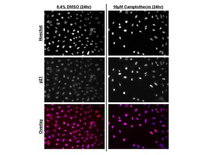
Immunofluorescence of camptothecin-treated A549 cells. A549 cells were plated on 96-well plates and treated with 10µM camptothecin or 0.4% DMSO control for 24 hours prior to fixation. Cell handling, fixation and immunostaining were performed using HCS234 kit reagents and protocols. Cells were imaged on a GE IN Cell Analyzer 1000 (3.4) at 20X objective magnification. Top and middle panels: Monochromatic images of Hoechst HCS Nuclear Stain and p21 fluorescence. Note the increase in p21 nuclear intensity following camptothecin treatment. Bottom panel: Fused images of Hoechst HCS Nuclear Stain (blue) and p21 fluorescence (red). Note that auto-contrasting in fused images may result in haziness due to low intensity signal contribution.
Immunofluorescence of camptothecin-treated A549 cells. A549 cells were plated on 96-well plates and treated with 10µM camptothecin or 0.4% DMSO control for 24 hours prior to fixation. Cell handling, fixation and immunostaining were performed using HCS234 kit reagents and protocols. Cells were imaged on a GE IN Cell Analyzer 1000 (3.4) at 20X objective magnification. Top and middle panels: Monochromatic images of Hoechst HCS Nuclear Stain and p21 fluorescence. Note the increase in p21 nuclear intensity following camptothecin treatment. Bottom panel: Fused images of Hoechst HCS Nuclear Stain (blue) and p21 fluorescence (red). Note that auto-contrasting in fused images may result in haziness due to low intensity signal contribution.


