The brain pattern of c-fos induction by two doses of amphetamine suggests different brain processing pathways and minor contribution of behavioural traits.
D Rotllant,C Márquez,R Nadal,A Armario
Neuroscience
168
2010
显示摘要
Previous studies have shown that amphetamine (AMPH) markedly activates dopaminergic projection areas, together with some important limbic nuclei. However, a global picture of the brain areas activated is lacking and the contribution of the dose of the drug and individual differences to this global brain activation is not known. In the present experiment, we studied in adult male rats the c-fos expression induced by two doses of AMPH (1.5 and 5 mg/kg sc) in a wide range of brain areas, and investigated the possible contribution of novelty-induced activity and anxiety traits. AMPH administration increased Fos+ neurons in an important number of telencephalic, diencephalic and brainstem areas. Interestingly, the ventral tegmental area (VTA) and the dorsal raphe nucleus were activated by the drug, but c-fos expression was restricted to non-dopaminergic and non-serotoninergic neurons, those activated in the VTA being predominantly GABAergic. The use of the factorial analysis, which grouped the areas in function of the correlation between the number of Fos+ neurons observed in each area, revealed three main factors, probably reflecting activation of various relatively independent brain circuits: the first included medial prefrontal cortex regions, most dorsal and ventral striatal subregions and VTA; the second, raphe nuclei; and the third, the different subdivisions of the paraventricular nucleus of the hypothalamus. Other areas such as the central amygdala did not group around any factor. The finding that an important number of activated areas grouped around specific factors is suggestive of activation of partially independent brain circuits. Surprisingly, a minor contribution of novelty-induced activity and anxiety traits on brain activation induced by AMPH was found. It is possible that normal variability in these traits is poorly related to the effects of AMPH or that c-fos expression is not a good tool to reveal such differences. | 20406670
 |
Immunohistochemical studies on phosphorylation of tyrosine hydroxylase in central catecholamine neurons using site- and phosphorylation state-specific antibodies.
Xu, Z Q, et al.
Neuroscience, 82: 727-38 (1998)
1998
显示摘要
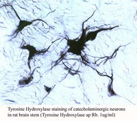
Antibodies raised to phosphorylated forms of tyrosine hydroxylase, the first and rate-limiting enzyme in the catecholamine biosynthesis, were applied in immunohistochemical studies on rat brain slices incubated in vitro with a phosphodiesterase inhibitor (3-isobutyl-1-methylxanthine, IBMX) and on forskolin on formalin-perfused rat brains. Four antisera/antibodies were used: polyclonal rabbit antisera to (i) tyrosine hydroxylase phosphorylated at serine 40 (THS40p antiserum), (ii) tyrosine hydroxylase phosphorylated at serine 19 (THS19p antiserum), (iii) the native enzyme (pan-tyrosine hydroxylase antiserum), and mouse monoclonal antibody to (iv) native tyrosine hydroxylase. In the in vitro studies THS40p-like immunoreactivity was not observed unless slices were treated with IBMX-forskolin after which a dense fibre network was found in the striatum, and immunoreactive cell bodies were found in the ventral mesencephalon, especially in the ventral tegmental area. Although these cells were pan-tyrosine hydroxylase-positive, several of them were not stained with the tyrosine hydroxylase-monoclonal antibody. Moreover, there was a marked reduction of tyrosine hydroxylase-monoclonal antibody-immunoreactive fibres in drug-treated slices, suggesting that this tyrosine hydroxylase-monoclonal antibody does not recognize the serine 40-phosphorylated form of tyrosine hydroxylase. Treated slices did not show any THS40p-immunoreactive cell bodies in the dopaminergic A11 cell group and only a few, weakly fluorescent neurons were observed in locus coeruleus. However, a sparse fibre plexus was observed in locus coeruleus, possibly reflecting epinephrine fibres. In the perfused brains THS40p-like immunoreactivity could be visualized in some dopamine neurons in the ventral mesencephalon, especially the A10 area, and in noradrenergic locus coeruleus neurons, whereas THS19p-like immunoreactivity was found in all catecholamine groups studied, similar to the results obtained with the pan-tyrosine hydroxylase antiserum and the tyrosine hydroxylase-monoclonal antibody. In forebrain areas known to be innervated by mesencephalic dopamine neurons, no THS40p-positive fibres were observed, whereas THS19p-immunoreactive fibres were found in subregions of the striatum, olfactory tubercle and nucleus accumbens, essentially overlapping with dopamine fibres previously shown to contain cholecystokinin-like immunoreactivity. The present results suggests that antibodies directed against phosphorylated forms of tyrosine hydroxylase can be used to evaluate the state of tyrosine hydroxylase phosphorylation in individual neuronal cell bodies and processes both in vitro and in vivo. | 9483531
 |
Antibodies to a segment of tyrosine hydroxylase phosphorylated at serine 40.
Goldstein, M, et al.
J. Neurochem., 64: 2281-7 (1995)
1995
显示摘要
A synthetic peptide corresponding to residues 32-47 of rat tyrosine hydroxylase (TH) was phosphorylated by protein kinase A at Ser40 and used to generate antibodies in rabbits. Reactivity of the anti-pTH32-47 antibodies with phospho- and dephospho-Ser40 forms of TH protein and peptide TH32-47 was compared with reactivity of antibodies to nonphosphorylated peptide and to native TH protein. In antibody-capture ELISAs, anti-pTH32-47 was more reactive with the phospho-TH than with the dephospho-TH forms. Conversely, antibodies against the nonphosphorylated peptide reacted preferentially with the dephospho-TH forms. In western blots, labeling of the approximately 60-kDa TH band by anti-pTH32-47 was readily detectable in lanes containing protein kinase A-phosphorylated native TH at 10-100 ng/lane. In blots of supernatants prepared from striatal synaptosomes, addition of a phosphatase inhibitor was necessary to discern labeling of the TH band with anti-pTH32-47. Similarly, anti-pTH32-47 failed to immunoprecipitate TH activity from supernatants prepared from untreated tissues, whereas prior treatment with either 8-bromoadenosine 3',5'-cyclic monophosphate or forskolin enabled removal of TH activity by anti-pTH32-47. Lastly, in immunohistochemical studies, anti-pTH32-47 selectively labeled catecholaminergic cells in tissue sections from perfusion-fixed rat brain. | 7722513
 |
Antibodies to a synthetic peptide corresponding to a Ser-40-containing segment of tyrosine hydroxylase: activation and immunohistochemical localization of tyrosine hydroxylase.
Lee, K Y, et al.
J. Neurochem., 53: 1238-44 (1989)
1989
显示摘要
A peptide corresponding to position 32-47 in tyrosine hydroxylase was synthesized (TH-16) and polyclonal antibodies against this peptide were raised in rabbits (anti-TH-16). The effects of anti-TH-16 on modulation of tyrosine hydroxylase activity were investigated. Anti-TH-16 enhanced the enzymatic activity in a concentration-dependent manner, and the antigen TH-16 inhibited the stimulatory activity of the antiserum in a concentration-dependent manner. The activated enzyme had a lower Km app for the cofactor 2-amino-4-hydroxy-6-methyl-5,6,7,8-tetrahydropterin and a higher Vmax app than the nonactivated enzyme. Anti-TH-16 was characterized further by its ability to immunoprecipitate the enzyme activity by labeling tyrosine hydroxylase after Western blotting and by immunohistochemical labeling of catecholaminergic neurons. Anti-TH-16 did not block activation of tyrosine hydroxylase by phosphorylation catalyzed by cyclic AMP-dependent protein kinase. Exposure of the enzyme to anti-TH-16 and subsequent phosphorylation of the enzyme resulted in a greater activation of the enzyme than the sum of activation produced by these two treatments separately. However, the activation was less than additive when the enzyme was first phosphorylated and subsequently exposed to anti-TH-16. The present study demonstrates the utility of anti-TH-16 in investigating the molecular aspects of the enzyme activation. | 2570128
 |


