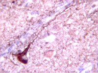
Pericyte loss influences Alzheimer-like neurodegeneration in mice.
Sagare, AP; Bell, RD; Zhao, Z; Ma, Q; Winkler, EA; Ramanathan, A; Zlokovic, BV
Nature communications
4
2932
2013
显示摘要
Pericytes are cells in the blood-brain barrier that degenerate in Alzheimer's disease (AD), a neurological disorder associated with neurovascular dysfunction, abnormal elevation of amyloid β-peptide (Aβ), tau pathology and neuronal loss. Whether pericyte degeneration can influence AD-like neurodegeneration and contribute to disease pathogenesis remains, however, unknown. Here we show that in mice overexpressing Aβ-precursor protein, pericyte loss elevates brain Aβ40 and Aβ42 levels and accelerates amyloid angiopathy and cerebral β-amyloidosis by diminishing clearance of soluble Aβ40 and Aβ42 from brain interstitial fluid prior to Aβ deposition. We further show that pericyte deficiency leads to the development of tau pathology and an early neuronal loss that is normally absent in Aβ-precursor protein transgenic mice, resulting in cognitive decline. Our data suggest that pericytes control multiple steps of AD-like neurodegeneration pathogenic cascade in Aβ-precursor protein-overexpressing mice. Therefore, pericytes may represent a novel therapeutic target to modify disease progression in AD. | 24336108
 |
Increased Tau Phosphorylation and Impaired Brain Insulin/IGF Signaling in Mice Fed a High Fat/High Cholesterol Diet.
Bhat, Narayan R and Thirumangalakudi, Lakshmi
J. Alzheimers Dis., (2013)
2013
显示摘要
Previous studies demonstrated that a high fat/high cholesterol (HFC) diet results in a loss of working memory in mice correlated with neuroinflammatory changes and increased AβPP processing (Thirumangalakudi et al. (2008) J Neurochem 106, 475-485). To further explore the nature of the molecular correlates of cognitive impairment, in this study, we examined changes in tau phosphorylation, insulin/IGF-1 signaling (IIS) including GSK3, and levels of specific synaptic proteins. Immunoblot analysis of hippocampal tissue from C57BL/6 mice fed HFC for 2 months with anti-phospho-tau (i.e., PHF1 and phospho-Thr-231 tau) antibodies demonstrated the presence of hyperphosphorylated tau. The tau phosphorylation correlated with activated GSK3, a prominent tau kinase normally kept inactive under the control of IIS. That IIS itself was impaired due to the hyperlipidemic diet was confirmed by a down-regulation of insulin receptor substrate-1 and phospho-Akt and levels. Although no significant changes in the levels of the pre-synaptic protein (i.e., synaptophysin) in response to HFC were apparent in immunoblot analysis, there was a clear down-regulation of the post-synaptic protein, PSD95, and drebrin, a dendritic spine-specific protein, indicative of altered synaptic plasticity. The results, in concert with previous findings with the same model, suggest that high dietary fat/cholesterol elicits brain insulin resistance and altered IIS leading to Alzheimer's disease-like cognitive impairment in 'normal' mice. | 23703152
 |
Dephosphorylation of Tau by protein phosphatase 5: impairment in Alzheimer's disease.
Liu, F., et al.
J. Biol. Chem., 280(3):1790-1796 (2005)
2005
| 15546861
 |
The retinoic acid and brain-derived neurotrophic factor differentiated SH-SY5Y cell line as a model for Alzheimer's disease-like tau phosphorylation.
Jämsä, A., et al.
Biochem. Biophys. Res. Comm., 319:993-1000 (2004)
2004
| 15184080
 |
O-GlcNAcylation regulates phosphorylation of tau: a mechanism involved in Alzheimer's disease.
Liu, Fei, et al.
Proc. Natl. Acad. Sci. U.S.A., 101: 10804-9 (2004)
2004
显示摘要
Microtubule-associated protein tau is abnormally hyperphosphorylated and aggregated into neurofibrillary tangles in brains of individuals with Alzheimer's disease (AD) and other tauopathies. Tau pathology is critical to pathogenesis and correlates to the severity of dementia. However, the mechanisms leading to abnormal hyperphosphorylation are unknown. Here, we demonstrate that human brain tau was modified by O-GlcNAcylation, a type of protein O-glycosylation by which the monosaccharide beta-N-acetylglucosamine (GlcNAc) attaches to serine/threonine residues via an O-linked glycosidic bond. O-GlcNAcylation regulated phosphorylation of tau in a site-specific manner both in vitro and in vivo. At most of the phosphorylation sites, O-GlcNAcylation negatively regulated tau phosphorylation. In an animal model of starved mice, low glucose uptake/metabolism that mimicked those observed in AD brain produced a decrease in O-GlcNAcylation and consequent hyperphosphorylation of tau at the majority of the phosphorylation sites. The O-GlcNAcylation level in AD brain extracts was decreased as compared to that in controls. These results reveal a mechanism of regulation of tau phosphorylation and suggest that abnormal hyperphosphorylation of tau could result from decreased tau O-GlcNAcylation, which probably is induced by deficient brain glucose uptake/metabolism in AD and other tauopathies. | 15249677
 |
Specific tau phosphorylation sites correlate with severity of neuronal cytopathology in Alzheimer's disease.
Augustinack, J.C., et al.
Acta Neuropathol., 103:26-35 (2002)
2002
| 11837744
 |
Site-specific phosphorylation of tau accompanied by activation of mitogen-activated protein kinase (MAPK) in brains of Niemann-Pick type C mice.
Sawamura, N, et al.
J. Biol. Chem., 276: 10314-9 (2001)
2001
显示摘要
Niemann-Pick type C (NPC) disease is characterized by an accumulation of cholesterol in most tissues and progressive neurodegeneration with the formation of neurofibrillary tangles. Neurofibrillary tangles are composed of paired helical filaments (PHF), a major component of which is the hyperphosphorylated tau. In this study we used NPC heterozygous and NPC homozygous mouse brains to investigate the molecular mechanism responsible for tauopathy in NPC. Immunoblot analysis using anti-tau antibodies (Tau-1, PHF-1, AT-180, and AT-100) revealed site-specific phosphorylation of tau at Ser-396 and Ser-404 in the brains of NPC homozygous mice. Mitogen-activated protein kinase, a potential serine kinase known to phosphorylate tau, was activated, whereas other serine kinases such as glycogen synthase kinase-3beta and cyclin-dependent kinase 5 were inactive. Morphological examination demonstrated that a number of neurons, the perikarya of which strongly immunostained with PHF-1, exhibited polymorphorous cytoplasmic inclusion bodies and multi-concentric lamellar-like bodies. Importantly, the accumulation of intracellular cholesterol in NPC mouse brains was determined to be a function of age. From these results we conclude that abnormal cholesterol metabolism due to the genetic mutation in NPC1 may be responsible for activation of the mitogen-activated protein kinase-signaling pathway and site-specific phosphorylation of tau in vivo, leading to tauopathy in NPC. | 11152466
 |
Expression of human apolipoprotein E4 in neurons causes hyperphosphorylation of protein tau in the brains of transgenic mice.
Tesseur,I., et al.
Am. J. Pathol., 156(3):951-964 (2000)
2000
| 10702411
 |
Phosphorylation of microtubule-associated protein tau is regulated by protein phosphatase 2A in mammalian brain. Implications for neurofibrillary degeneration in Alzheimer's disease.
Gong, C.X., et al.
J. Biol. Chem., 275(8):5535-5544 (2000)
2000
| 10681533
 |
Tau is phosphorylated by GSK-3 at several sites found in Alzheimer disease and its biological activity markedly inhibited only after it is prephosphorylated by A-kinase.
Wang, J.Z., et al.
FEBS Lett., 436(1):28-34 (1998)
1998
| 9771888
 |


