Millipore Sigma Vibrant Logo
Attention: We have moved. Merck Millipore products are no longer available for purchase on MerckMillipore.com.Learn More

ST1042 Anti-PKD2 Rabbit pAb

ST1042
Purchase on Sigma-Aldrich

概述

Replacement Information

重要规格表

Species ReactivityHostAntibody Type
HRbPolyclonal Antibody

Products

产品目录编号包装 数量 / 包装
ST1042-50UGCN 塑胶安瓿;塑胶针药瓶 50 μg
Description
OverviewRecognizes the ~105 kDa PKD2 protein in HEK293 cells.
Catalogue NumberST1042
Brand Family Calbiochem®
SynonymsAnti-Protein Kinase D2
Application Data
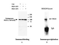
Panel A: Detection of human PKD2 by immunoblotting. Samples: Whole cell lysate (30 µg) from untransfected HEK 293 cells (Ctrl, left lane), HEK 293 cells transfected with PKD3-GFP fusion protein (~136 kDa, middle lane), or HEK 293 cells transfected with PKD1-GFP fusion protein (~136 kDa, right lane). Primary antibody: Anti-PKD2 Rabbit pAb (Cat. No. ST1042) (1:1000) Detection: chemiluminescence. Panel B: Detection of human PKD2 by immunoprecipitation, followed by immunoblotting. Sample: Whole cell lysate (200 µg protein) from HEK 293 cells. Antibody for IP: Anti-PKD2 Rabbit pAb (Cat. No. ST1042) (1:100). Immunoblotting conditions: same as above.
References
ReferencesRey, O., et al. 2003. Biochem. Biophys. Res. Commun. 302, 817.
Sturany, S., et al. 2002. J. Biol. Chem. 277, 29431.
Sturany, S., et al. 2001. J. Biol. Chem. 276, 3310.
Product Information
FormLiquid
FormulationIn PBS.
Positive controlHEK 293 cells
Preservative≤0.1% sodium azide
Quality LevelMQ100
Applications
Key Applications Immunoblotting (Western Blotting)
Immunoprecipitation
Application NotesImmunoblotting (1:500-1:5000)
Immunoprecipitation (1-4 µg/mg lysate)
Application CommentsThis antibody does not cross-react with PKD1 or PKD3. Antibody should be titrated for optimal use in individual systems.
Biological Information
Immunogena synthetic peptide corresponding to amino acids near the C-terminus of human PKD2
ImmunogenHuman
HostRabbit
IsotypeIgG
Species Reactivity
  • Human
Antibody TypePolyclonal Antibody
Concentration Label Please refer to vial label for lot-specific concentration
Physicochemical Information
Dimensions
Materials Information
Toxicological Information
Safety Information according to GHS
Safety Information
Product Usage Statements
Storage and Shipping Information
Ship Code Blue Ice Only
Toxicity Highly Toxic
Storage +2°C to +8°C
Do not freeze Ok to freeze
Special InstructionsFollowing initial use, aliquot and freeze (-20°C) for long-term storage. Avoid freeze/thaw.
Packaging Information
Transport Information
Supplemental Information
Specifications
Global Trade Item Number
产品目录编号 GTIN
ST1042-50UGCN 04055977224368

Documentation

Anti-PKD2 Rabbit pAb MSDS

职位

物料安全数据表 (MSDS) 

Anti-PKD2 Rabbit pAb 分析证书

标题批号
ST1042

参考

参考信息概述
Rey, O., et al. 2003. Biochem. Biophys. Res. Commun. 302, 817.
Sturany, S., et al. 2002. J. Biol. Chem. 277, 29431.
Sturany, S., et al. 2001. J. Biol. Chem. 276, 3310.
数据表

Note that this data sheet is not lot-specific and is representative of the current specifications for this product. Please consult the vial label and the certificate of analysis for information on specific lots. Also note that shipping conditions may differ from storage conditions.

Revision13-March-2009 JSW
SynonymsAnti-Protein Kinase D2
ApplicationImmunoblotting (1:500-1:5000)
Immunoprecipitation (1-4 µg/mg lysate)
Application Data
Panel A: Detection of human PKD2 by immunoblotting. Samples: Whole cell lysate (30 µg) from untransfected HEK 293 cells (Ctrl, left lane), HEK 293 cells transfected with PKD3-GFP fusion protein (~136 kDa, middle lane), or HEK 293 cells transfected with PKD1-GFP fusion protein (~136 kDa, right lane). Primary antibody: Anti-PKD2 Rabbit pAb (Cat. No. ST1042) (1:1000) Detection: chemiluminescence. Panel B: Detection of human PKD2 by immunoprecipitation, followed by immunoblotting. Sample: Whole cell lysate (200 µg protein) from HEK 293 cells. Antibody for IP: Anti-PKD2 Rabbit pAb (Cat. No. ST1042) (1:100). Immunoblotting conditions: same as above.
DescriptionImmunoaffinity purified rabbit polyclonal antibody. Recognizes the ~105 kDa PKD2 protein.
BackgroundProtein kinase D2 (PKD2) is a serine/threonine protein kinase and a member of PKD family, which also includes PKD/PKD1/PKCµ and PKD3/PKCν. PKDs are a newly identified protein kinase family with structural, enzymological and regulatory properties different from protein kinase C (PKC) family. PKDs participate in the signal transduction pathway and are potential regulators of cell proliferation and apoptosis. They are stimulated by diacyl-glycerol (DAG), and are substrates for classical PKCs like PKCα, PKCε and PKCη. Therefore, PKD stimulation by DAG is partially mediated through PKCs. PKD2 mRNA is widely expressed in human and murine tissue and codes for a ~105 kDa protein. It is stimulated by both phorbol ester and growth factors and is autophosphorylated upon stimulation. PKD2 is predominantly located in the cytoplasm and, as opposed to other members of the PKD family, does not accumulate in the nucleus upon induction. This suggests that PKD2 mediates physiological responses different from other members of the PKD family.
HostRabbit
Immunogen speciesHuman
Immunogena synthetic peptide corresponding to amino acids near the C-terminus of human PKD2
IsotypeIgG
Specieshuman
Positive controlHEK 293 cells
FormLiquid
FormulationIn PBS.
Concentration Label Please refer to vial label for lot-specific concentration
Preservative≤0.1% sodium azide
CommentsThis antibody does not cross-react with PKD1 or PKD3. Antibody should be titrated for optimal use in individual systems.
Storage +2°C to +8°C
Do Not Freeze Ok to freeze
Special InstructionsFollowing initial use, aliquot and freeze (-20°C) for long-term storage. Avoid freeze/thaw.
Toxicity Highly Toxic
ReferencesRey, O., et al. 2003. Biochem. Biophys. Res. Commun. 302, 817.
Sturany, S., et al. 2002. J. Biol. Chem. 277, 29431.
Sturany, S., et al. 2001. J. Biol. Chem. 276, 3310.