Millipore Sigma Vibrant Logo
Attention: We have moved. Merck Millipore products are no longer available for purchase on MerckMillipore.com.Learn More

386032 Anti-Hsp70 Mouse mAb (C92F3A-5)

386032
Purchase on Sigma-Aldrich

概述

Replacement Information

重要规格表

Species ReactivityHostAntibody Type
A Broad Range Of SpeciesMMonoclonal Antibody

Products

产品目录编号包装 数量 / 包装
386032-50UGCN 塑胶安瓿;塑胶针药瓶 50 μg
Description
OverviewRecognizes the ~70 kDa Hsp70 protein. Does not cross-react with Hsc70.
Catalogue Number386032
Brand Family Calbiochem®
SynonymsAnti-Heat Shock Protein 70
Application Data
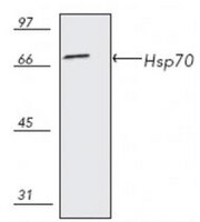
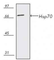
Detection of mouse Hsp70 by immunoblotting immunoblotting. Sample: Whole cell lysate from L929 cells. Primary antibody: Anti-Hsp70 Mouse mAb (C92F3A-5) (Cat. No. 386032) (1:1000). Detection: chemiluminescence.

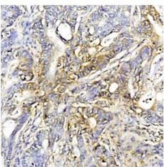
Detection of human Hsp70 by staining paraffin sections. Sample: Human colon cancer tissue. Primary antibody: Anti-Hsp70 Mouse mAb (C92F3A-5) (Cat. No. 386032) (1:50). Detection: colorimetric (DAB).

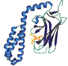
Substrate binding of Hsp70 in complex with a substrate peptide.
References
ReferencesHang, H., and Fox, M.H. 1995. Cytometry 19, 119.
Kilgore, J.L., et al. 1994. J. Appl. Physiol. 76, 589.
Heufelder, A.E., et al. 1992. J. Clin. Endocrinol. Metab. 74, 724.
Gower, D.J., et al. 1989. J. Neurosurg. 70, 605.
Milarski, K., et al. 1989.J. Cell Biol. 108, 413.
Vass, K., et al. 1988. Acta Neuropathologica 77, 128.
Welch, W.J., and Suhan, J.P. 1986. J. Cell Biol. 103, 2035.
Product Information
FormLiquid
FormulationIn PBS, 50% glycerol, pH 7.2.
Positive controlL929 cells, Human colon cancer tissue
Preservative≤0.1% sodium azide
Quality LevelMQ100
Applications
Key Applications Enzyme-Linked Immunosorbent Assay
Flow Cytometry
Frozen Sections
Immunoblotting (Western Blotting)
Immunocytochemistry
Immunoprecipitation
Paraffin Sections
Application NotesELISA (see comments)
Flow Cytometry (see comments)
Frozen Sections (5 µg/ml)
Immunoblotting (1 µg/ml)
Immunocytochemistry (5 µg/ml)
Immunoprecipitation (see comments)
Paraffin Sections (5 µg/ml)
Application CommentsDoes not cross-react with Hsc70. Because Hsp70 is not expressed constitutively in most cells, this antibody is suitable for ascertaining whether a stress-response has occurred in the cell. Increased levels of Hsp70 expression occurs following stress even in cells that constitutively express Hsp70. This antibody has also been reported to work for ELISA, flow cytometry, and immunoprecipitation. Variables associated with assay conditions will dictate the optimal working dilution.
Biological Information
ImmunogenHsp70 from HeLa cells
ImmunogenHuman
CloneC92F3A-5
HostMouse
IsotypeIgG₁
Species Reactivity
  • A Broad Range Of Species
Antibody TypeMonoclonal Antibody
Concentration Label Please refer to vial label for lot-specific concentration
Physicochemical Information
Dimensions
Materials Information
Toxicological Information
Safety Information according to GHS
Safety Information
Product Usage Statements
Storage and Shipping Information
Ship Code Blue Ice Only
Toxicity Standard Handling
Storage -20°C
Avoid freeze/thaw Avoid freeze/thaw
Do not freeze Ok to freeze
Special InstructionsFollowing iniital thaw, aliquot and freeze (-20°C).
Packaging Information
Transport Information
Supplemental Information
Specifications
Global Trade Item Number
产品目录编号 GTIN
386032-50UGCN 04055977189889

Documentation

Anti-Hsp70 Mouse mAb (C92F3A-5) MSDS

职位

物料安全数据表 (MSDS) 

Anti-Hsp70 Mouse mAb (C92F3A-5) 分析证书

标题批号
386032

参考

参考信息概述
Hang, H., and Fox, M.H. 1995. Cytometry 19, 119.
Kilgore, J.L., et al. 1994. J. Appl. Physiol. 76, 589.
Heufelder, A.E., et al. 1992. J. Clin. Endocrinol. Metab. 74, 724.
Gower, D.J., et al. 1989. J. Neurosurg. 70, 605.
Milarski, K., et al. 1989.J. Cell Biol. 108, 413.
Vass, K., et al. 1988. Acta Neuropathologica 77, 128.
Welch, W.J., and Suhan, J.P. 1986. J. Cell Biol. 103, 2035.
数据表

Note that this data sheet is not lot-specific and is representative of the current specifications for this product. Please consult the vial label and the certificate of analysis for information on specific lots. Also note that shipping conditions may differ from storage conditions.

Revision10-June-2009 JSW
SynonymsAnti-Heat Shock Protein 70
ApplicationELISA (see comments)
Flow Cytometry (see comments)
Frozen Sections (5 µg/ml)
Immunoblotting (1 µg/ml)
Immunocytochemistry (5 µg/ml)
Immunoprecipitation (see comments)
Paraffin Sections (5 µg/ml)
Application Data
Detection of mouse Hsp70 by immunoblotting immunoblotting. Sample: Whole cell lysate from L929 cells. Primary antibody: Anti-Hsp70 Mouse mAb (C92F3A-5) (Cat. No. 386032) (1:1000). Detection: chemiluminescence.

Detection of human Hsp70 by staining paraffin sections. Sample: Human colon cancer tissue. Primary antibody: Anti-Hsp70 Mouse mAb (C92F3A-5) (Cat. No. 386032) (1:50). Detection: colorimetric (DAB).

Substrate binding of Hsp70 in complex with a substrate peptide.
DescriptionMouse monoclonal antibody purified by ion-exchange chromatography. Recognizes the ~70 kDa Hsp70 protein.
BackgroundThe Hsp70 family consists of multiple members, all of which bind ATP in vitro, that are localized within different intracellular compartments. Hsp70 is not usually expressed under normal growth conditions, but is expressed at high levels in cells experiencing stress. It is found in the cytosol, nucleus, and nucleolus.
HostMouse
Immunogen speciesHuman
ImmunogenHsp70 from HeLa cells
CloneC92F3A-5
IsotypeIgG₁
Speciesa broad range of species
Positive controlL929 cells, Human colon cancer tissue
FormLiquid
FormulationIn PBS, 50% glycerol, pH 7.2.
Concentration Label Please refer to vial label for lot-specific concentration
Preservative≤0.1% sodium azide
CommentsDoes not cross-react with Hsc70. Because Hsp70 is not expressed constitutively in most cells, this antibody is suitable for ascertaining whether a stress-response has occurred in the cell. Increased levels of Hsp70 expression occurs following stress even in cells that constitutively express Hsp70. This antibody has also been reported to work for ELISA, flow cytometry, and immunoprecipitation. Variables associated with assay conditions will dictate the optimal working dilution.
Storage Avoid freeze/thaw
-20°C
Do Not Freeze Ok to freeze
Special InstructionsFollowing iniital thaw, aliquot and freeze (-20°C).
Toxicity Standard Handling
ReferencesHang, H., and Fox, M.H. 1995. Cytometry 19, 119.
Kilgore, J.L., et al. 1994. J. Appl. Physiol. 76, 589.
Heufelder, A.E., et al. 1992. J. Clin. Endocrinol. Metab. 74, 724.
Gower, D.J., et al. 1989. J. Neurosurg. 70, 605.
Milarski, K., et al. 1989.J. Cell Biol. 108, 413.
Vass, K., et al. 1988. Acta Neuropathologica 77, 128.
Welch, W.J., and Suhan, J.P. 1986. J. Cell Biol. 103, 2035.