Millipore Sigma Vibrant Logo
Attention: We have moved. Merck Millipore products are no longer available for purchase on MerckMillipore.com.Learn More

NE1015 Anti-Glial Fibrillary Acidic Protein Cocktail Mouse mAb (SMI-22)

NE1015
Purchase on Sigma-Aldrich

概述

Replacement Information

重要规格表

Species ReactivityHostAntibody Type
B, Ca, Ch, Gp, H, M, Porcine, R, ShMMonoclonal Antibody

Products

产品目录编号包装 数量 / 包装
NE1015-100ULCN 塑胶安瓿;塑胶针药瓶 100 ul
Description
OverviewRecognizes ~50 kDa glial fibrillary acidic protein (GFAP) in human and bovine cytoskeletal preparations.
Catalogue NumberNE1015
Brand Family Calbiochem®
SynonymsAnti-GFAP Cocktail
Application Data
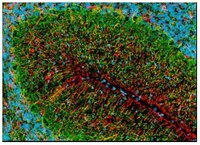
Detection of rat glial fibrillary acidic protein by staining frozen sections. Sample: Rat brain. Primary antibody: Anti-GFAP Cocktail Mouse mAb (SMI-22) (Cat. No. NE1015) (1:1000). Detection: fluorescence (red) with Hoechst 33342 counterstain.

Primary mixed glial cultures were stained with anti-mouse GFAP (EMD Millipore, Cat. No. MAB360) and a DyLight™488 conjugated goat anti-mouse secondary Antibody. The nucleus was counterstained with DAPI. The image was captured using Olympus BX51 with an exposure time of 982 millisec for green channel and 126 millisec for DAPI channel. Magnification is 10X without any correction on Gain/Offset/Bad pixel. Courtesy of Jose Shinsmon, Immunology, Dept of Pathology, Faculty of Medicine and Health Sciences, UPM,Serdang 43400.
References
ReferencesVick, W.W., et al. 1987. Acta. Cytol. 31, 816.
McLendon R.E., et al. 1986. J. Neuropathol. Exp. Neurol. 45, 692.
Pegram, C.N., et al. 1985. Neurochem. Pathol. 3, 119.
Product Information
FormLiquid
FormulationUndiluted ascites.
Positive controlAstrocytes or cytoskeletal preparations
Preservative≤ 0.1% sodium azide
Quality LevelMQ100
Applications
Key Applications Enzyme-Linked Immunosorbent Assay
Frozen Sections
Immunoblotting (Western Blotting)
Immunocytochemistry
Paraffin Sections
Application NotesELISA (1:1000)
Frozen Sections (1:1000, see comments)
Immunoblotting (1:1000)
Immunocytochemistry (1:1000, see comments)
Paraffin Sections (1:1000, trypsin or heat pre-treatment required)
Application CommentsThis cocktail is derived from the Bigner-Eng clones MAb1B4, MAb2E1, and MAb4A11 and provides a means for more comprehensive detection of astrocytomas than each clone alone. Each component is specific for GFAP and stains astrocytes and astrocytic processes as well as Bergman glia. Recognizes both anaplastic and reactive astrocytes by immunocytochemical staining. Does not recognize metastatic tumors and brain tumors of non-astrocytic origin, including medulloblastomas, meningiomas, choroid plexus papillomas, and schwannomas. For staining paraffin sections it is recommended that de-paraffinized sections be treated with 0.1% trypsin in 50 mM Tris-HCl, pH 7.6 for 20-30 min at 37°C or boiled in Tris-buffered saline, pH 9.0 for 15 min to expose the epitope. For immunocytochemistry or staining frozen sections, post-fixation in cold methanol or methanol/hydrogen peroxide for 10 min is required for access to the astrocytes in the sample. Antibody should be titrated for optimal results in individual systems.
Biological Information
Immunogenpurified bovine GFAP protein
ImmunogenBovine
CloneSMI-22
HostMouse
IsotypeIgG2b
Species Reactivity
  • Bovine
  • Canine
  • Chicken
  • Guinea Pig
  • Human
  • Mouse
  • Porcine
  • Rat
  • Sheep
Antibody TypeMonoclonal Antibody
Physicochemical Information
Dimensions
Materials Information
Toxicological Information
Safety Information according to GHS
Safety Information
Product Usage Statements
Storage and Shipping Information
Ship Code Dry Ice Only
Toxicity Standard Handling
Storage -20°C
Avoid freeze/thaw Avoid freeze/thaw
Do not freeze Ok to freeze
Special InstructionsUpon initial thaw, aliquot and freeze (-20°C).
Packaging Information
Transport Information
Supplemental Information
Specifications
Global Trade Item Number
产品目录编号 GTIN
NE1015-100ULCN 04055977209853

Documentation

Anti-Glial Fibrillary Acidic Protein Cocktail Mouse mAb (SMI-22) MSDS

职位

物料安全数据表 (MSDS) 

Anti-Glial Fibrillary Acidic Protein Cocktail Mouse mAb (SMI-22) 分析证书

标题批号
NE1015

参考

参考信息概述
Vick, W.W., et al. 1987. Acta. Cytol. 31, 816.
McLendon R.E., et al. 1986. J. Neuropathol. Exp. Neurol. 45, 692.
Pegram, C.N., et al. 1985. Neurochem. Pathol. 3, 119.
数据表

Note that this data sheet is not lot-specific and is representative of the current specifications for this product. Please consult the vial label and the certificate of analysis for information on specific lots. Also note that shipping conditions may differ from storage conditions.

Revision01-October-2007 RFH
SynonymsAnti-GFAP Cocktail
ApplicationELISA (1:1000)
Frozen Sections (1:1000, see comments)
Immunoblotting (1:1000)
Immunocytochemistry (1:1000, see comments)
Paraffin Sections (1:1000, trypsin or heat pre-treatment required)
Application Data
Detection of rat glial fibrillary acidic protein by staining frozen sections. Sample: Rat brain. Primary antibody: Anti-GFAP Cocktail Mouse mAb (SMI-22) (Cat. No. NE1015) (1:1000). Detection: fluorescence (red) with Hoechst 33342 counterstain.

Primary mixed glial cultures were stained with anti-mouse GFAP (EMD Millipore, Cat. No. MAB360) and a DyLight™488 conjugated goat anti-mouse secondary Antibody. The nucleus was counterstained with DAPI. The image was captured using Olympus BX51 with an exposure time of 982 millisec for green channel and 126 millisec for DAPI channel. Magnification is 10X without any correction on Gain/Offset/Bad pixel. Courtesy of Jose Shinsmon, Immunology, Dept of Pathology, Faculty of Medicine and Health Sciences, UPM,Serdang 43400.
DescriptionMouse monoclonal antibody cocktail that contains a mixture of 3 antibodies supplied as undiluted ascites. Recognizes the ~50 kDa glial fibrillary acidic protein.
BackgroundGlial fibrillary acidic protein (GFAP) is an intermediate filament protein found only in glial cells or cells of glial origin. It can be detected in astrocytes and certain other astroglia in the CNS, in satellite cells in peripheral ganglia, and in non-myelinating Schwann cells in peripheral nerves. GFAP is upregulated and expressed at high levels in astrocytes in many damage and disease states. It is also often highly expressed in neural stem cells and many types of brain tumors.
HostMouse
Immunogen speciesBovine
Immunogenpurified bovine GFAP protein
CloneSMI-22
IsotypeIgG2b
Speciesbovine, canine, chicken, guinea pig, human, mouse, porcine, rat, sheep
Positive controlAstrocytes or cytoskeletal preparations
FormLiquid
FormulationUndiluted ascites.
Preservative≤ 0.1% sodium azide
CommentsThis cocktail is derived from the Bigner-Eng clones MAb1B4, MAb2E1, and MAb4A11 and provides a means for more comprehensive detection of astrocytomas than each clone alone. Each component is specific for GFAP and stains astrocytes and astrocytic processes as well as Bergman glia. Recognizes both anaplastic and reactive astrocytes by immunocytochemical staining. Does not recognize metastatic tumors and brain tumors of non-astrocytic origin, including medulloblastomas, meningiomas, choroid plexus papillomas, and schwannomas. For staining paraffin sections it is recommended that de-paraffinized sections be treated with 0.1% trypsin in 50 mM Tris-HCl, pH 7.6 for 20-30 min at 37°C or boiled in Tris-buffered saline, pH 9.0 for 15 min to expose the epitope. For immunocytochemistry or staining frozen sections, post-fixation in cold methanol or methanol/hydrogen peroxide for 10 min is required for access to the astrocytes in the sample. Antibody should be titrated for optimal results in individual systems.
Storage Avoid freeze/thaw
-20°C
Do Not Freeze Ok to freeze
Special InstructionsUpon initial thaw, aliquot and freeze (-20°C).
Toxicity Standard Handling
ReferencesVick, W.W., et al. 1987. Acta. Cytol. 31, 816.
McLendon R.E., et al. 1986. J. Neuropathol. Exp. Neurol. 45, 692.
Pegram, C.N., et al. 1985. Neurochem. Pathol. 3, 119.