Millipore Sigma Vibrant Logo
Attention: We have moved. Merck Millipore products are no longer available for purchase on MerckMillipore.com.Learn More

ABE1957 Anti-CENP-C Antibody

ABE1957
100 µL  
Purchase on Sigma-Aldrich

特卖

概述

Replacement Information

重要规格表

Species ReactivityKey ApplicationsHostFormatAntibody Type
HWB, ICCRbSerumPolyclonal Antibody
Description
Catalogue NumberABE1957
DescriptionAnti-CENP-C Antibody
Alternate Names
  • Centromere protein C
  • CENP-C
  • CENP-C 1
  • Centromere autoantigen C
  • Centromere protein C 1
  • Interphase centromere complex protein 7
Background InformationCentromere protein C (UniProt Q03188; also known as CENP-C, CENP-C 1, Centromere autoantigen C, Centromere protein C 1, Interphase centromere complex protein 7) is encoded by the CENPC (also known as CENPC1, ICEN7) gene (Gene ID 1060) in human. Kinetochores assemble on centromeres to bind spindle microtubules and mediate chromosome segregation during cell division. CENP-C is a subunit of constitutive centromere-associated network (CCAN) that acts as the centromere-kinetochore interface. CCAN is a large complex composed of at least 16 different centromeric proteins (CENPs). CENP-C plays an important role in kinetochore assembly downstream of CENP-A. The PEST domain in the N-terminal half of CENP-C interacts directly with the CCAN subcomplex CENP-HIKM (CENP-H, CENP-K, CENP-I, and CENP-M) and CENP-C depletion is shown to cause mislocalization of CENP-T and the CENP-HIKM complex subunits in HeLa cells.
References
Product Information
FormatSerum
PresentationRabbit polyclonal antibody serum with 0.05% sodium azide.
Quality LevelMQ100
Applications
Application
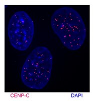
Anti-CENP-C Antibody, Cat. No. ABE1957, is a highly specific rabbit polyclonal antibody that targets CENP-C and has been tested in Immunocytochemistry and Western Blotting.
Key Applications
  • Western Blotting
  • Immunocytochemistry
Application NotesImmunocytochemistry Analysis: A 1:10,000 dilution from a representative lot immunostained kinetochores in chromosome spreads prepared from interphase DLD-1 human colorectal adenocarcinoma cells hypotonically swollen and fixed with 4% formaldehyde.

Immunocytochemistry Analysis: A representative lot was affinity purified and detected a time-dependent loss of kinetochores CENP-C immunoreactivity in 4% formaldehyde-fixed, 0.5% Triton X-100-permeabilized HeLa cells following CENP-C shRNA induction (Falk, S.J., et al. (2015). Science. 348(6235):699-703).

Immunocytochemistry Analysis: A representative lot was affinity purified and immunostained kinetochores by indirect fluorescence staining of chromosome spreads prepared from patient-derived, PD-NC4 chromosome variant harboring fibroblasts hypotonically swollen and fixed with 4% formaldehyde (Bassett, E.A., et al. (2010). J. Cell Biol. 190(2):177-185).

Western Blotting Analysis: A representative lot was affinity purified and detected a time-dependent CENP-C level in HeLa cells following CENP-C shRNA induction (Falk, S.J., et al. (2015). Science. 348(6235):699-703).
Biological Information
ImmunogenGST-tagged recombinant human CENP-C N-terminal fragment.
EpitopeN-terminus
ConcentrationPlease refer to lot specific datasheet.
HostRabbit
SpecificityThis rabbit polyclonal antiserum targets the N-terminal region of human CENP-C present in both spliced isoforms reported by UniProt (Q03188). Specificity was demonstrated by a reducion of target band detection (Western blotting) and kinetochores staining (chromsome spreads fluoresence staining) following cellular CENP-C shRNA induction (Falk, S.J., et al. (2015). Science. 348(6235):699-703).
Species Reactivity
  • Human
Species Reactivity NoteHuman.
Antibody TypePolyclonal Antibody
Entrez Gene Number
Gene Symbol
  • CENPC
  • CENPC1
  • ICEN7
Purification MethodUnpurified.
UniProt Number
Molecular Weight~140 kDa observed. 106.8 kDa (isoform 1) and 61.54 kDa (isoform 2) calculated. The larger-than-calculated target band size is consistent with that reported in the literature (Earnshaw, W.C., et al. (1989). Chromosoma. 98(1):1-12). Uncharacterized bands may be observed in some lysate(s).
Physicochemical Information
Dimensions
Materials Information
Toxicological Information
Safety Information according to GHS
Safety Information
Product Usage Statements
Quality AssuranceEvaluated by Western Blotting in DLD-1 cell lysate.

Western Blotting Analysis: A 1:5,000 dilution of this antiserum detected CENP-C in 50,000 cell equivalent of DLD-1 human colorectal adenocarcinoma cell lysate.
Usage Statement
  • Unless otherwise stated in our catalog or other company documentation accompanying the product(s), our products are intended for research use only and are not to be used for any other purpose, which includes but is not limited to, unauthorized commercial uses, in vitro diagnostic uses, ex vivo or in vivo therapeutic uses or any type of consumption or application to humans or animals.
Storage and Shipping Information
Storage ConditionsStable for 1 year at -20°C from date of receipt.
Handling Recommendations: Upon receipt and prior to removing the cap, centrifuge the vial and gently mix the solution. Aliquot into microcentrifuge tubes and store at -20°C. Avoid repeated freeze/thaw cycles, which may damage IgG and affect product performance.
Packaging Information
Material Size100 µL
Transport Information
Supplemental Information
Specifications
Global Trade Item Number
产品目录编号 GTIN
ABE1957 04054839090103