Millipore Sigma Vibrant Logo
Attention: We have moved. Merck Millipore products are no longer available for purchase on MerckMillipore.com.Learn More

MABT365 Anti-CEACAM1 Antibody, clone 11-1H

MABT365
100 µg  
Purchase on Sigma-Aldrich

特卖

概述

Replacement Information

重要规格表

Species ReactivityKey ApplicationsHostFormatAntibody Type
RWB, IHC, ELISA, FC, IPMPurifiedMonoclonal Antibody
Description
Catalogue NumberMABT365
DescriptionAnti-CEACAM1 Antibody, clone 11-1H
Alternate Names
  • CEACAM1
  • Carcinoembryonic antigen-related cell adhesion molecule 1
  • ATP-dependent taurocolate-carrier protein
  • Cell-CAM 105
  • C-CAM 105
  • Ecto-ATPase
  • GP110
  • pp120
  • CD66a
Background InformationThe carcinoembryonic antigen- (CEA-) related cell adhesion molecules (CEACAMs) constitute a 12-member subgroup of the CEA family of immunoglobulin-related proteins first described in 1965 (PMID 4953873). CEACAMs are reported to participate in diverse physiological processes, including cell adhesion, differentiation, proliferation, and survival, as well as in carcinogenesis and bacterial pathogenesis. CEACAM molecules from neighbouring cells can interact via their respective extracellular N-terminal IgV-like domain and mediate cell-cell adhesion through trans-oligomerization. CEACAM molecules within the same cell can also undergo transmembrane domain-mediated cis-oligomerization, an event important for sustaining downstream cellular signaling. Athough CEACAM1 is shown to utilize its cytoplasmic domain for transducing cellular signaling, not all CEACAM members are expressed with a significant cytoplasmic domain, and others (CEACAM5/6/7/8) are GPI-anchored without even a transmembrane domain. (PMID 4735478). Rodent Ceacam1 (also known as C-CAM 105, Cell-CAM 105, Ecto-ATPase, GP110, pp120; UniProt P16573) is encoded by the Ceacam1 gene (NCBI Gene ID 81613). As is the case with its human counterpart (P13688), there exisit a long (canonical; 519aa) and a short (458aa) form with a reduced cytoplamic domain as a result of alternate splicing. An additional 10 splice variants are also reported for human CEACAM1. Clone 11-1H does not cross-react with human CEACAM1/3/4/5/6/7/8 or mouse CEACAM1/2.
References
Product Information
FormatPurified
PresentationPurified mouse monoclonal IgG1κ in buffer containing 0.1 M Tris-Glycine (pH 7.4), 150 mM NaCl with 0.05% sodium azide.
Quality LevelMQ300
Applications
ApplicationAnti-CEACAM1 Antibody, clone 11-1H is an antibody against CEACAM1 for use in Western Blotting, Immunohistochemistry, ELISA, Flow Cytometry and Immunoprecipitation.
Key Applications
  • Western Blotting
  • Immunohistochemistry
  • ELISA
  • Flow Cytometry
  • Immunoprecipitation
Application NotesImmunohistochemistry Analysis: A 1:50-250 dilution from a representative lot detected CEACAM1 in rat liver, lung and kidney tissue.
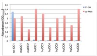
ELISA Analysis: A representative lot detected CEACAM1 in CHO-cells expressing CEACAM1s (Dr. B. Singer, University Duisburg-Essen).
Flow Cytometry Analysis: A representative lot detected CEACAM1 in CHO-ratCEACAM1 transfected cells (Dr. B. Singer, University Duisburg-Essen).
Immunoprecipitation Analysis: A representative lot immunoprecipitated CEACAM1 in epithelial rat bladder carcinoma cell line known to express rat CEACAM1 (Dr. B. Singer, University Duisburg-Essen).
Western Blotting Analysis: A representative lot detected CEACAM1 in CHO-ratCEACAM1 and CHO-rat CEACAM1dN lysate (Dr. B. Singer, University Duisburg-Essen).
Immunohistochemistry Analysis: A representative lot detected CEACAM1 in rat prostate tissue (Dr. B. Singer, University Duisburg-Essen).
Biological Information
ImmunogenRecombinant protein corresponding to the extracellular, excluding N domain of rat CEACAM1.
EpitopeExtracellular, excluding N domain
Clone11-1H
ConcentrationPlease refer to lot specific datasheet.
HostMouse
IsotypeIgG1κ
Species Reactivity
  • Rat
Antibody TypeMonoclonal Antibody
Entrez Gene Number
Gene Symbol
  • Ceacam1
Purification MethodProtein G Purified
UniProt Number
Molecular Weight~80 kDa observed. The calculated molecular weight of this protein is 55 kDa, but can be observed at ~80-140 kDa due to high glycosylation.
Physicochemical Information
Dimensions
Materials Information
Toxicological Information
Safety Information according to GHS
Safety Information
Product Usage Statements
Quality AssuranceEvaluated by Western Blotting in rat liver tissue lysate.

Western Blotting Analysis: 0.5 µg/mL of this antibody detected CEACAM1 in 10 µg of rat liver tissue lysate.
Usage Statement
  • Unless otherwise stated in our catalog or other company documentation accompanying the product(s), our products are intended for research use only and are not to be used for any other purpose, which includes but is not limited to, unauthorized commercial uses, in vitro diagnostic uses, ex vivo or in vivo therapeutic uses or any type of consumption or application to humans or animals.
Storage and Shipping Information
Storage ConditionsStable for 1 year at 2-8°C from date of receipt.
Packaging Information
Material Size100 µg
Transport Information
Supplemental Information
Specifications
Global Trade Item Number
产品目录编号 GTIN
MABT365 04055977162790