Millipore Sigma Vibrant Logo
Attention: We have moved. Merck Millipore products are no longer available for purchase on MerckMillipore.com.Learn More

MABE1068 Anti-APE/Ref-1 Antibody, clone 13B8E5C2

MABE1068
100 μg  
Purchase on Sigma-Aldrich

特卖

概述

Replacement Information

重要规格表

Species ReactivityKey ApplicationsHostFormatAntibody Type
M, H, RICC, WB, IH(P)MPurifiedMonoclonal Antibody
Description
Catalogue NumberMABE1068
DescriptionAnti-APE/Ref-1 Antibody, clone 13B8E5C2
Alternate Names
  • DNA-(apurinic or apyrimidinic site) lyase
  • AP endonuclease 1
  • APE-1
  • APEN
  • APEX nuclease
  • Apurinic-apyrimidinic endonuclease 1
  • Redox factor-1
  • REF-1
Background InformationDNA-(apurinic or apyrimidinic site) lyase (EC 4.2.99.18; UniProt P27695; also known as AP endonuclease 1, APE-1, APEN, APEX nuclease, Apurinic-apyrimidinic endonuclease 1, Redox factor-1, REF-1) is encoded by the APEX1 (also known as APE, APE1, APEX, APX, HAP1, REF1) gene (Gene ID 328) in human. APE/Ref-1 mediates the cleavage of an apurinic/apyrimidinic (AP) site in DNA generated by DNA glycosylases during base excision repair (BER) in both nucleus and mitochondria. APE/Ref-1 also functions as a redox factor (ref) responsible for maintaining several transcription factors (TFs) in an active reduced state for DNA binding, including AP-1, NF-κB, HIF-1α, CREB, TP53. Elevated levels of APE1 are associated with resistance to chemotherapyand poor prognosis in various cancers. APE/Ref-1 is synthesized as a 318-a.a. protein whose initiation methionine (Met1) is removed posttranslationally to yield the 317-a.a. form with a nuclear localization sequence (NLS; a.a. 8-13) and a nuclear export signal (NES; a.a. 64-80) sequence to allow its shuttle between the cytoplasm and nucleus. The 317-a.a. form can be further processed by a mitochondria-associated N-terminal peptidase into a mitochrondrial form (mtAPE; a.a. 32-318) that lacks NLS and contains only the NES and the mitochondrial targeting sequence (MTS; a.a. 289-318).
References
Product Information
FormatPurified
PresentationPurified mouse IgG2a in buffer containing 0.1 M Tris-Glycine (pH 7.4), 150 mM NaCl with 0.05% sodium azide.
Quality LevelMQ100
Applications
ApplicationAnti-APE/Ref-1, clone 13B8E5C2, Cat. No. MABE1068, is a highly specific mouse monoclonal antibody, that targets APE1 and has been tested in Immunocytochemistry, Immunohistochemistry (Paraffin), and Western Blotting.
Key Applications
  • Immunocytochemistry
  • Western Blotting
  • Immunohistochemistry (Paraffin)
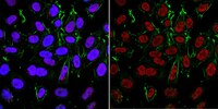
Application NotesImmunocytochemistry Analysis: A 1:200 dilution from a representative lot detected both cytosolic and nuclear APE/Ref-1 immunoreactivity in 4% paraformaldehyde-fixed, 0.3% Triton X-100-permeabilized HeLa cells.

Immunohistochemistry Analysis: A 1:1,000 dilution from a representative lot detected nuclear APE/Ref-1 immunoreactivity in rat kidney, and human placenta & small intestine tissue sections.

Western Blotting Analysis: 0.5 µg/mL from a representative lot detected APE/Ref-1 in 10 µg of Raji cell lysate.

Immunocytochemistry Analysis: A representative lot detected both cytosolic and nuclear APE/Ref-1 immunoreactivity in HeyC2 and HEY human ovarian cancer cell lines (Moore, D.H., et al. (2000). Clin. Cancer Res. 6(2):602-609).

Immunohistochemistry Analysis: A representative lot detected varying tissue distribution and cellular localization of APE/Ref-1 immunoreactivity among various 4% formaldehyde-fixed, paraffin-embedded human ovarian tissue samples, including ovarian endometriosis, serous cystadenoma , serous carcinoma, ands well as normal ovarian tissues (Moore, D.H., et al. (2000). Clin. Cancer Res. 6(2):602-609).

Western Blotting Analysis: A representative lot detected an upregulated cytosolic APE/Ref-1 level in HeyC2 than HEY cells, while similar nuclear APE/Ref-1 level was detected in these two human ovarian cancer cell lines (Moore, D.H., et al. (2000). Clin. Cancer Res. 6(2):602-609).
Biological Information
ImmunogenRecombinant full-length human APE/Ref-1.
Clone13B8E5C2
ConcentrationPlease refer to lot specific datasheet.
HostMouse
SpecificityClone 13B8E5C2 detected both cytosolic and nuclear APE/Ref-1 by Immunocytochemistry, Immunohistochemistry, and Western blotting (Moore, D.H., et al. (2000). Clin. Cancer Res. 6(2):602-609).
IsotypeIgG2aκ
Species Reactivity
  • Mouse
  • Human
  • Rat
Species Reactivity NoteHuman, Mouse, Rat.
Antibody TypeMonoclonal Antibody
Entrez Gene Number
Gene Symbol
  • APEX1
  • APE
  • APE1
  • APEX
  • APX
  • HAP1
  • REF1
Purification MethodProtein G purified.
UniProt Number
Molecular Weight~35-39 kDa observed. 35.42 kDa (Met1 removed; a.a. 2-318) and 32.24 kDa (Mitochondrial form; a.a. 32-318) calculated. The broad banding pattern is consistent with the detection of both mitochondrial and non-mitochondrial forms with varying degrees of posttranslational modifications (lysine acetylation, threonine phosphorylation, cysteine nitrosylation). Uncharacterized bands may be observed in some lysate(s).
Physicochemical Information
Dimensions
Materials Information
Toxicological Information
Safety Information according to GHS
Safety Information
Product Usage Statements
Quality AssuranceEvaluated by Western Blotting in HepG2 cell lysate.

Western Blotting Analysis: 0.5 µg/mL of this antibody detected APE/Ref-1 in 10 µg of HepG2 cell lysate.
Usage Statement
  • Unless otherwise stated in our catalog or other company documentation accompanying the product(s), our products are intended for research use only and are not to be used for any other purpose, which includes but is not limited to, unauthorized commercial uses, in vitro diagnostic uses, ex vivo or in vivo therapeutic uses or any type of consumption or application to humans or animals.
Storage and Shipping Information
Storage ConditionsStable for 1 year at 2-8°C from date of receipt.
Packaging Information
Material Size100 μg
Transport Information
Supplemental Information
Specifications
Global Trade Item Number
产品目录编号 GTIN
MABE1068 04054839090264