Millipore Sigma Vibrant Logo

LMR001 Cell Capture Imaging Reagent

LMR001
8 mL  
Ricerca del prezzo in corso...
Non è stato possibile trovare il prezzo
La quantità minima deve essere un multiplo di
Maximum Quantity is
Al termine dell'ordine Maggiori informazioni
Lei ha salvato ()
 
Richiedi il prezzo
Disponibilità limitata
A magazzino 
Fuori produzione
Disponibili quantità limitate
La disponibilità deve essere confermata
    Prodotti rimanenti: riceverà un nostro avviso
      Prodotti rimanenti: riceverà un nostro avviso
      Will advise
      Contatti il Servizio Clienti
      Contact Customer Service


      Contatti il Servizio Clienti

      Panoramica

      Replacement Information
      Description
      Catalogue NumberLMR001
      DescriptionCell Capture Imaging Reagent
      Alternate Names
      • Photopolymeric hydrogel
      Background InformationCell Capture Imaging Reagent is a specially formulated photopolymeric hydrogel thin film that is optically transparent and is permeable to antibodies and other immunocytochemistry reagents. It allows cells to be physically constrained at the bottom of standard imaging microtiter without any cell loss. It is particularly suitable for use in experiments where the cell population is less than 10,000. Cells captured in this hydrogel can be easily micrographed regardless of the staining reagents. This polymeric hydrogel displays sufficient mechanical strength to withstand routine pipetting and is dense enough for cells to sink to the imaging surface.
      References
      Product Information
      FormatLiquid Reagent
      PresentationClear viscous liquid.
      Applications
      ApplicationCell Capture Imaging Reagent is a specially formulated polymeric hydrogel that allows cells to be constrained at the bottom of microtiter plates for subsequent imaging.
      Key Applications
      • Immunocytochemistry
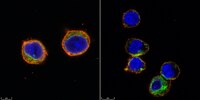
      Application NotesImmunocytochemistry Analysis: Use of this reagent with an antibody detected Jurkat cells.
      Biological Information
      ConcentrationPlease refer to lot specific datasheet.
      Species Reactivity Note.
      UniProt Number
      Molecular WeightN/A
      Physicochemical Information
      Dimensions
      Materials Information
      Toxicological Information
      Safety Information according to GHS
      Safety Information
      Product Usage Statements
      Quality Assurance1. Wash cells with 1x PBS twice
      2. Re-suspend cells using PBS to concentration < 1 x 10⁶ cells / mL
      3. In 384- Imaging well-plate, Add 40 µL of cell suspension per well. Number of cells will vary depending on user preference.
      4. Add 6.5 µL of Cell Capture Imaging Reagent. Do not mix. Gently dispense using pipette.

      NOTE:
      This ratio of cell volume to Cell Capture Imaging Reagent reagent volume is valid when using 384 Corning High Content microplate with glass bottom plates (Catalog Number 4581). If using a different plate with different dimension this ratio must be recalculated:
      Example:
      • 384 Corning High Content microplate with glass bottom plate (4581): .0625 cm2
      • 96 A/2 Corning High Content microplate (4580): .1586 cm2
      • Ratio: .1586/.0625= 2.54 Conversion Factor
      • Cells: 40ul*2.54= 101.5ul of Cells
      • Cell Capture Imaging Reagent: 6.5ul*2.54= 16.5 of Cell Capture Imaging Reagent

      5. Centrifuge well-plate for 5 mins @ 500 g
      6. Expose to UV-light for 15 seconds on ChemiDoc with standard UV wavelength, 302 nm
      NOTE:
      Exposure time may need to adjust based on UV intensity. UV light must come from the bottom of the well. To optimize UV exposure, a short time course with the correct ratio of PBS to Cell Capture Imaging Reagent can be used. To visualize formation, hold 96 well plate up to light to distinguish a slight yellow color and by taking a pipet tip and remove the gel plug from well. Use the shortest amount of UV exposure possible.
      Alternatively, to optimize UV exposure, take any cells resuspended in PBS with the appropriate amount of Cell Capture Imaging Reagent and expose to UV. Start with the shortest amount of time followed by steps 7-9 below and by a 10 min DAPI stain and washing with PBS. Visualize on microscope to make sure cell retention is high. This same procedure can also be used to determine a suitable number of cells to dispense into well.
      7. Remove supernatant from each well by flicking plate
      8. Add 1x PBS each well. Take caution not to poke the bottom layer. Discard supernatant by flicking the plate.
      9. Repeat step 8 four times.
      10. Cells are ready for immunocytochemistry.
      All immunocytochemistry steps can be performed in the well with Cell Capture Imaging Reagent encapsulated cells. Be sure to keep wells hydrated with PBS or buffer at all times.
      Usage Statement
      • Unless otherwise stated in our catalog or other company documentation accompanying the product(s), our products are intended for research use only and are not to be used for any other purpose, which includes but is not limited to, unauthorized commercial uses, in vitro diagnostic uses, ex vivo or in vivo therapeutic uses or any type of consumption or application to humans or animals.
      Storage and Shipping Information
      Storage ConditionsStable for 1 year at 2-8°C from date of receipt.
      Packaging Information
      Material Size8 mL
      Transport Information
      Supplemental Information
      Specifications
      Global Trade Item Number
      Numero di catalogo GTIN
      LMR001 04061841037675