Millipore Sigma Vibrant Logo

OP15L Anti-c-ErbB2/c-Neu (Ab-3) Mouse mAb (3B5)

View Products on Sigmaaldrich.com
OP15L
Visualizza prezzi e disponibilità

Panoramica

Replacement Information

Tabella delle specifiche principali

Species ReactivityHostAntibody Type
H, MMMonoclonal Antibody

Prezzi e disponibilità

Numero di catalogo DisponibilitàConfezionamento Qtà/conf Prezzo Quantità
OP15L-100UG
Verifica della disponibilità in corso...
Disponibilità limitata
A magazzino 
Fuori produzione
Disponibili quantità limitate
La disponibilità deve essere confermata
    Prodotti rimanenti: riceverà un nostro avviso
      Prodotti rimanenti: riceverà un nostro avviso
      Will advise
      Contatti il Servizio Clienti
      Contact Customer Service

      Bottiglia di vetro 100 μg
      Ricerca del prezzo in corso...
      Non è stato possibile trovare il prezzo
      La quantità minima deve essere un multiplo di
      Maximum Quantity is
      Al termine dell'ordine Maggiori informazioni
      Lei ha salvato ()
       
      Richiedi il prezzo
      Description
      OverviewRecognizes the ~185 kDa c-ErbB2/c-Neu protein in HEK293 cells.
      Catalogue NumberOP15L
      Brand Family Calbiochem®
      SynonymsAnti-ErbB2, Anti-Erythroblastosis Virus, Anti-Human Epidermal Growth Factor, Anti-HER2, Anti-Neu
      Application Data
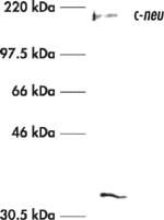
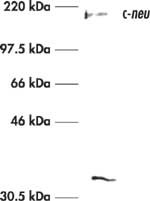
      Detection of human c-ErbB2/c-Neu by immunoblotting. Sample: Whole cell lysate from HEK293 cells. Primary antibody: Anti-c-ErbB2/c-Neu (Ab-3) Mouse mAb (3B5) (Cat. No. OP15L) (1 µg/ml). Detection: chemiluminescence.
      References
      ReferencesMamcs, J.R., et al. 1994. Ann. Surg. 219, 332.
      Tetu, B., et al. 1994. Cancer 73, 2359.
      DiFiore, P.P., et al. 1987. Science 237, 178.
      Slamon, D.J., et al. 1987. Science 235, 177.
      Varley, J.M., et al. 1987. Oncogene 1, 423.
      Bargmann, C.I., et al. 1986. Nature 319, 226.
      Yamamoto, T., et al. 1986. Nature 319, 230.
      Blick, M., et al. 1984. Blood 64, 1234.
      Schwab, M., et al. 1984. Cold Spring Harbor Laboratory 2, 215.
      Product Information
      FormLyophilized
      FormulationLyophilized from 20 mM ammonium bicarbonate Buffer, 100 µg FAF-BSA.
      Negative controlHepG2 cells or normal skin
      Positive controlSK-BR-3 cells or breast carcinoma tissue
      PreservativeNone
      Quality LevelMQ100
      Applications
      Application ReferencesParaffin Sections Meissner, K., et al. 1990. Am. J. Pathol. 137, 1305. Van De Vijver, J.J., et al. 1988. New Engl. J. Med. 319, 1239. Original Clone Van De Vijver, J.J., et al. 1988. New Engl. J. Med. 319, 1239. Immunohistochemistry, Immunoblotting Zhau, H.E., et al. 1990. Mol. Carcinogen. 3, 254. Immunoprecipitation Van De Vijver, J.J., et al. 1988. New Engl. J. Med. 319, 1239.
      Key Applications Frozen Sections
      Immunoblotting (Western Blotting)
      Immunofluorescence
      Immunoprecipitation
      Paraffin Sections
      Application NotesFrozen Sections (2.5 µg/ml)
      Immunoblotting (2.5 µg/ml, see application references)
      Immunofluorescence (2.5 µg/ml)
      Immunoprecipitation (1 µg per sample, see application references)
      Paraffin Sections (2.5 µg/ml, pressure cooker pre-treatment required, see application references)
      Application CommentsFor paraffin sections, we recommend pre-treating with a pressure cooker, but trypsin and heat will also work well. Including 0.05% saponin during staining of paraffin sections may help reduce background. In one study ductal carcinomas in situ displaying large-cell, comedo growth type, were stained while none of 16 ductal carcinomas in situ of small-cell, papillary, or cribriform growth type were stained by this antibody. Antibody should be titrated for optimal results in individual systems.
      Biological Information
      Immunogena synthetic peptide (TAENPEYLGLDVPV) corresponding to amino acids 1242-1255 from the C-terminal domain of human c-ErbB2/c-Neu
      ImmunogenHuman
      Clone3B5
      HostMouse
      IsotypeIgG₁
      Species Reactivity
      • Human
      • Mouse
      Antibody TypeMonoclonal Antibody
      Physicochemical Information
      Dimensions
      Materials Information
      Toxicological Information
      Safety Information according to GHS
      Safety Information
      Product Usage Statements
      Storage and Shipping Information
      Ship Code Ambient Temperature Only
      Toxicity Standard Handling
      Storage +2°C to +8°C
      Do not freeze Ok to freeze
      Special InstructionsReconstitue the lyophilized antibody with sterile PBS, pH 7.4, or sterile 20 mM Tris-saline (20 mM Tris containing 0.15 M NaCl), pH 7.4, to yield a final concentration of 100 µg/ml. Lyophilized antibodies should be reconstituted at 4°C with occasional gentle mixing for at least 2 h. Store at 4°C until reconstituted, then store in aliquots at -20°C or at 4°C with 0.1% azide. Freezing of aliquots is best for long term storage of reconstituted product; repetitive freezing and thawing should be avoided.
      Packaging Information
      Transport Information
      Supplemental Information
      Specifications
      Global Trade Item Number
      Numero di catalogo GTIN
      OP15L-100UG 04055977224641

      Documentation

      Anti-c-ErbB2/c-Neu (Ab-3) Mouse mAb (3B5) MSDS

      Titolo

      Scheda di sicurezza (MSDS) 

      Anti-c-ErbB2/c-Neu (Ab-3) Mouse mAb (3B5) Certificati d'Analisi

      TitoloNumero di lotto
      OP15L

      Riferimenti bibliografici

      Panoramica delle referenze
      Mamcs, J.R., et al. 1994. Ann. Surg. 219, 332.
      Tetu, B., et al. 1994. Cancer 73, 2359.
      DiFiore, P.P., et al. 1987. Science 237, 178.
      Slamon, D.J., et al. 1987. Science 235, 177.
      Varley, J.M., et al. 1987. Oncogene 1, 423.
      Bargmann, C.I., et al. 1986. Nature 319, 226.
      Yamamoto, T., et al. 1986. Nature 319, 230.
      Blick, M., et al. 1984. Blood 64, 1234.
      Schwab, M., et al. 1984. Cold Spring Harbor Laboratory 2, 215.

      Citazioni

      Titolo
    • Rita Nahta, et al. (2005) Insulin-like growth factor-I receptor/human epidermal growth factor receptor 2 heterodimerization contributes to trastuzumab resistance of breast cancer cells. Cancer Research 65, 11118-11128.
    • Scheda tecnica

      Note that this data sheet is not lot-specific and is representative of the current specifications for this product. Please consult the vial label and the certificate of analysis for information on specific lots. Also note that shipping conditions may differ from storage conditions.

      Revision02-October-2007 RFH
      SynonymsAnti-ErbB2, Anti-Erythroblastosis Virus, Anti-Human Epidermal Growth Factor, Anti-HER2, Anti-Neu
      ApplicationFrozen Sections (2.5 µg/ml)
      Immunoblotting (2.5 µg/ml, see application references)
      Immunofluorescence (2.5 µg/ml)
      Immunoprecipitation (1 µg per sample, see application references)
      Paraffin Sections (2.5 µg/ml, pressure cooker pre-treatment required, see application references)
      Application Data
      Detection of human c-ErbB2/c-Neu by immunoblotting. Sample: Whole cell lysate from HEK293 cells. Primary antibody: Anti-c-ErbB2/c-Neu (Ab-3) Mouse mAb (3B5) (Cat. No. OP15L) (1 µg/ml). Detection: chemiluminescence.
      DescriptionPurified mouse monoclonal antibody generated by immunizing BALB/c mice with the specified immunogen and fusing with Sp2/0.Ag14 cells (see application references). Recognizes the ~185 kDa c-ErbB2/c-Neu protein.
      BackgroundAmplification of the human proto-oncogene c-neu has been reported to occur in approximately 30% of breast carcinomas and measurement of the amplification has been proposed as a useful predictor of disease prognosis. Gene amplification and the resulting overexpression of proto-oncogene encoded p185c-neu is thought to transform cells by chronically stimulating signal transduction pathways, and in model systems overexpression of human c-neu induces cellular transformation. In the rat model system, a point mutation has been located within the transmembrane domain of the c-neu oncogene. neu (from neuroglioblastomas) is also referred to as erbB-2 (from erythroblastosis virus) and HER2 (for Human Epidermal Growth Factor Receptor) and was the second of four members of the EGF receptor family to be described.
      HostMouse
      Immunogen speciesHuman
      Immunogena synthetic peptide (TAENPEYLGLDVPV) corresponding to amino acids 1242-1255 from the C-terminal domain of human c-ErbB2/c-Neu
      Clone3B5
      IsotypeIgG₁
      Specieshuman, mouse
      Positive controlSK-BR-3 cells or breast carcinoma tissue
      Negative controlHepG2 cells or normal skin
      FormLyophilized
      FormulationLyophilized from 20 mM ammonium bicarbonate Buffer, 100 µg FAF-BSA.
      PreservativeNone
      CommentsFor paraffin sections, we recommend pre-treating with a pressure cooker, but trypsin and heat will also work well. Including 0.05% saponin during staining of paraffin sections may help reduce background. In one study ductal carcinomas in situ displaying large-cell, comedo growth type, were stained while none of 16 ductal carcinomas in situ of small-cell, papillary, or cribriform growth type were stained by this antibody. Antibody should be titrated for optimal results in individual systems.
      Storage +2°C to +8°C
      Do Not Freeze Ok to freeze
      Special InstructionsReconstitue the lyophilized antibody with sterile PBS, pH 7.4, or sterile 20 mM Tris-saline (20 mM Tris containing 0.15 M NaCl), pH 7.4, to yield a final concentration of 100 µg/ml. Lyophilized antibodies should be reconstituted at 4°C with occasional gentle mixing for at least 2 h. Store at 4°C until reconstituted, then store in aliquots at -20°C or at 4°C with 0.1% azide. Freezing of aliquots is best for long term storage of reconstituted product; repetitive freezing and thawing should be avoided.
      Toxicity Standard Handling
      ReferencesMamcs, J.R., et al. 1994. Ann. Surg. 219, 332.
      Tetu, B., et al. 1994. Cancer 73, 2359.
      DiFiore, P.P., et al. 1987. Science 237, 178.
      Slamon, D.J., et al. 1987. Science 235, 177.
      Varley, J.M., et al. 1987. Oncogene 1, 423.
      Bargmann, C.I., et al. 1986. Nature 319, 226.
      Yamamoto, T., et al. 1986. Nature 319, 230.
      Blick, M., et al. 1984. Blood 64, 1234.
      Schwab, M., et al. 1984. Cold Spring Harbor Laboratory 2, 215.
      Citation
    • Rita Nahta, et al. (2005) Insulin-like growth factor-I receptor/human epidermal growth factor receptor 2 heterodimerization contributes to trastuzumab resistance of breast cancer cells. Cancer Research 65, 11118-11128.
    • Application referencesParaffin Sections Meissner, K., et al. 1990. Am. J. Pathol. 137, 1305. Van De Vijver, J.J., et al. 1988. New Engl. J. Med. 319, 1239. Original Clone Van De Vijver, J.J., et al. 1988. New Engl. J. Med. 319, 1239. Immunohistochemistry, Immunoblotting Zhau, H.E., et al. 1990. Mol. Carcinogen. 3, 254. Immunoprecipitation Van De Vijver, J.J., et al. 1988. New Engl. J. Med. 319, 1239.

      Prodotti e applicazioni correlate

      Related Products

      Numero di catalogo Descrizione  
      OP15 Anti-c-ErbB2/c-Neu (Ab-3) Mouse mAb (3B5) Prezzi e disponibilità

      Categorie

      Life Science Research > Antibodies and Assays > Primary Antibodies