


386032 Sigma-AldrichAnti-Hsp70 Mouse mAb (C92F3A-5)
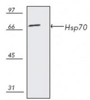

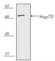
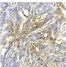
Anti-Hsp70, mouse monoclonal, clone C92F3A-5, recognizes the ~70 kDa Hsp70. Does not cross-react with Hsc70. It is validated for use in ELISA, FC, WB, ICC, IP & IHC (frozen and paraffin sections).
More>> Anti-Hsp70, mouse monoclonal, clone C92F3A-5, recognizes the ~70 kDa Hsp70. Does not cross-react with Hsc70. It is validated for use in ELISA, FC, WB, ICC, IP & IHC (frozen and paraffin sections). Less<<Anti-Hsp70 Mouse mAb (C92F3A-5): MSDS (material safety data sheet) o SDS, certificato d’analisi (CoA) e certificato di qualità (CoQ), dossier, brochure e altri documenti disponibili.
Sinonimi: Anti-Heat Shock Protein 70
Prodotti consigliati
Panoramica
| Replacement Information |
|---|
Tabella delle specifiche principali
| Species Reactivity | Host | Antibody Type |
|---|---|---|
| A Broad Range Of Species | M | Monoclonal Antibody |
Prezzi e disponibilità
| Numero di catalogo | Disponibilità | Confezionamento | Qtà/conf | Prezzo | Quantità | |
|---|---|---|---|---|---|---|
| 386032-50UG |
|
Fiala di plastica | 50 μg |
|
— |
| Product Information | |
|---|---|
| Form | Liquid |
| Formulation | In PBS, 50% glycerol, pH 7.2. |
| Positive control | L929 cells, Human colon cancer tissue |
| Preservative | ≤0.1% sodium azide |
| Quality Level | MQ100 |
| Physicochemical Information |
|---|
| Dimensions |
|---|
| Materials Information |
|---|
| Toxicological Information |
|---|
| Safety Information according to GHS |
|---|
| Safety Information |
|---|
| Product Usage Statements |
|---|
| Packaging Information |
|---|
| Transport Information |
|---|
| Supplemental Information |
|---|
| Specifications |
|---|
| Global Trade Item Number | |
|---|---|
| Numero di catalogo | GTIN |
| 386032-50UG | 04055977189889 |
Documentation
Anti-Hsp70 Mouse mAb (C92F3A-5) MSDS
| Titolo |
|---|
Anti-Hsp70 Mouse mAb (C92F3A-5) Certificati d'Analisi
| Titolo | Numero di lotto |
|---|---|
| 386032 |
Riferimenti bibliografici
| Panoramica delle referenze |
|---|
| Hang, H., and Fox, M.H. 1995. Cytometry 19, 119. Kilgore, J.L., et al. 1994. J. Appl. Physiol. 76, 589. Heufelder, A.E., et al. 1992. J. Clin. Endocrinol. Metab. 74, 724. Gower, D.J., et al. 1989. J. Neurosurg. 70, 605. Milarski, K., et al. 1989.J. Cell Biol. 108, 413. Vass, K., et al. 1988. Acta Neuropathologica 77, 128. Welch, W.J., and Suhan, J.P. 1986. J. Cell Biol. 103, 2035. |