


MABT51 Sigma-AldrichAnti-Galectin-3 Antibody, clone M3/38


Anti-Galectin-3 Antibody, clone M3/38 is an antibody against Galectin-3 for use in WB, IP, IH & IC.
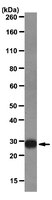
More>> Anti-Galectin-3 Antibody, clone M3/38 is an antibody against Galectin-3 for use in WB, IP, IH & IC. Less<<Anti-Galectin-3 Antibody, clone M3/38: MSDS (material safety data sheet) o SDS, certificato d’analisi (CoA) e certificato di qualità (CoQ), dossier, brochure e altri documenti disponibili.
Panoramica
| Replacement Information |
|---|
Tabella delle specifiche principali
| Species Reactivity | Key Applications | Host | Format | Antibody Type |
|---|---|---|---|---|
| H, M | WB, IP, IHC, ICC | R | Purified | Monoclonal Antibody |
| References |
|---|
| Product Information | |
|---|---|
| Format | Purified |
| Control |
|
| Presentation | Purified rat monoclonal IgG2aκ in buffer containing 0.1 M Tris-Glycine (pH 7.4), 150 mM NaCl with 0.05% sodium azide. |
| Quality Level | MQ100 |
| Physicochemical Information |
|---|
| Dimensions |
|---|
| Materials Information |
|---|
| Toxicological Information |
|---|
| Safety Information according to GHS |
|---|
| Safety Information |
|---|
| Storage and Shipping Information | |
|---|---|
| Storage Conditions | Stable for 1 year at 2-8°C from date of receipt. |
| Packaging Information | |
|---|---|
| Material Size | 50 µg |
| Transport Information |
|---|
| Supplemental Information |
|---|
| Specifications |
|---|
| Global Trade Item Number | |
|---|---|
| Numero di catalogo | GTIN |
| MABT51 | 04053252290985 |
Documentation
Anti-Galectin-3 Antibody, clone M3/38 MSDS
| Titolo |
|---|
Anti-Galectin-3 Antibody, clone M3/38 Certificati d'Analisi
| Titolo | Numero di lotto |
|---|---|
| Anti-Galectin-3, clone M3/38 | 2476943 |
| Anti-Galectin-3, clone M3/38 | 2474212 |
| Anti-Galectin-3, clone M3/38 - 2135135 | 2135135 |
| Anti-Galectin-3, clone M3/38 - 1991862 | 1991862 |
| Anti-Galectin-3, clone M3/38 - 2225189 | 2225189 |
| Anti-Galectin-3, clone M3/38 - 2327014 | 2327014 |
| Anti-Galectin-3, clone M3/38 - 2531557 | 2531557 |
| Anti-Galectin-3, clone M3/38 - 3174817 | 3174817 |
| Anti-Galectin-3, clone M3/38 - 3524845 | 3524845 |
| Anti-Galectin-3, clone M3/38 - 3613019 | 3613019 |