Expansion of vitiligo lesions is associated with reduced epidermal CDw60 expression and increased expression of HLA-DR in perilesional skin.
Le Poole, IC; Stennett, LS; Bonish, BK; Dee, L; Robinson, JK; Hernandez, C; Hann, SK; Nickoloff, BJ
The British journal of dermatology
149
739-48
2003
Mostra il sommario
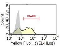
Detection of CDw60 in skin is representative of ganglioside D3 expression. This ganglioside is expressed primarily by melanocytes, and is of interest as a membrane antigen targeted by immunotherapy for melanoma patients. Expression of CDw60 by keratinocytes is defined by the presence of T-helper cell (Th)1 vs. Th2 cytokines, and can serve as a sentinel molecule to characterize an ongoing skin immune response.These immunobiological characteristics have provided the incentive to study the expression of CDw60 in the context of progressive vitiligo.Frozen sections were obtained from control skin and from vitiligo lesions and immunostained to show CDw60. Cells were cultured, their CDw60 expression studied and ribonuclease protection assays run to detect cytokine mRNA.Resistance to cytokine-mediated regulation of CDw60 expression was demonstrated in vitro by melanocytes, which appeared capable of generating autocrine and paracrine regulatory molecules supporting CDw60 expression. Induction of CDw60 expression was inhibited by antibodies to interleukin (IL)-4, suggesting that this cytokine was responsible, at least in part, for melanocyte-induced CDw60 expression. Marginal skin from patients with progressive generalized vitiligo consistently showed a reduction in epidermal CDw60 expression alongside elevated human leucocyte associated antigen (HLA)-DR expression at the margin. It thus appears that inflammatory infiltrates present in marginal skin generate type 1 rather than type 2 cytokines, supportive of a cell-mediated autoimmune response.These results support an active role of melanocytes within the skin immune system, and associate their loss in generalized vitiligo with a cell-mediated immune response mediated by type 1 cytokines. | 14616364
 |
Activation pathways of synovial T lymphocytes. Expression and function of the UM4D4/CDw60 antigen.
Fox, DA; Millard, JA; Kan, L; Zeldes, WS; Davis, W; Higgs, J; Emmrich, F; Kinne, RW
The Journal of clinical investigation
86
1124-36
1990
Mostra il sommario
Accumulating evidence implicates a central role for synovial T cells in the pathogenesis of rheumatoid arthritis, but the activation pathways that drive proliferation and effector function of these cells are not known. We have recently generated a novel monoclonal antibody against a rheumatoid synovial T cell line that recognizes an antigen termed UM4D4 (CDw60). This antigen is expressed on a minority of peripheral blood T cells, and represents the surface component of a distinct pathway of human T cell activation. The current studies were performed to examine the expression and function of UM4D4 on T cells obtained from synovial fluid and synovial membranes of patients with rheumatoid arthritis and other forms of inflammatory joint disease. The UM4D4 antigen is expressed at high surface density on about three-fourths of synovial fluid T cells and on a small subset of synovial fluid natural killer cells; in synovial tissue it is present on more than 90% of T cells in lymphoid aggregates, and on approximately 50% of T cells in stromal infiltrates In addition, UM4D4 is expressed in synovial tissue on a previously undescribed population of HLA-DR/DP-negative non-T cells with a dendritic morphology. Anti-UM4D4 was co-mitogenic for both RA and non-RA synovial fluid mononuclear cells, and induced IL-2 receptor expression. The UM4D4/CDw60 antigen may represent a functional activation pathway for synovial compartment T cells, which could play an important role in the pathogenesis of inflammatory arthritis. | 2212003
 |
A novel pathway of human T lymphocyte activation. Identification by a monoclonal antibody generated against a rheumatoid synovial T cell line.
Higgs, JB; Zeldes, W; Kozarsky, K; Schteingart, M; Kan, L; Bohlke, P; Krieger, K; Davis, W; Fox, DA
Journal of immunology (Baltimore, Md. : 1950)
140
3758-65
1987
Mostra il sommario
Substantial evidence indicates that compartmentalized infiltrates of T lymphocytes are central to the pathogenesis of autoimmune diseases such as rheumatoid arthritis, but the mechanisms by which such cells become activated remain unknown. To define surface components of activation pathways important in the function of these cells, we have generated mAb against a rheumatoid synovial T cell line. One such antibody, termed anti-UM4D4, reacts with an Ag, termed UM4D4, which is strongly expressed on most rheumatoid synovial T cell lines and clones, and on a subset of peripheral blood T cells, resting or activated. Anti-UM4D4 is mitogenic in soluble form for PBMC and certain T cell clones, and is comitogenic with the phorbol ester PMA for purified resting T lymphocytes. These functional effects are similar to those previously observed with antibodies to epitopes of CD2 and CD3, surface Ag involved in two well defined pathways of human T cell activation. Binding of anti-UM4D4 to T cells is not, however, blocked by antibodies directed at various epitopes of CD2 and CD3. Moreover, UM4D4 does not comodulate with CD3, and is expressed on a T cell line that lacks CD2, CD3, and CD28. The data, therefore, indicate that anti-UM4D4 identifies a T cell activation pathway, distinct from those previously described, that could play a role in the pathogenesis of T cell-mediated autoimmune diseases. | 2836500
 |


