Aberrant expression of G1-phase cell cycle regulators in flat and exophytic adenomas of the human colon.
Bartkova, J; Thullberg, M; Slezak, P; Jaramillo, E; Rubio, C; Thomassen, LH; Bartek, J
Gastroenterology
120
1680-8
2001
Mostra il sommario
The G1/S-phase controlling mechanism known as the RB pathway is commonly deregulated in human malignancies. Here, the abundance and localization of key components of the retinoblastoma (RB) pathway were determined in exophytic and flat colorectal adenomas.Samples of normal colonic mucosa (n = 41) and flat (n = 45) and exophytic (n = 26) adenomas were examined immunohistochemically using antibodies to cyclins D1, D2, D3, cyclin-dependent kinase (CDK) 4, retinoblastoma protein (pRB), and the CDK inhibitors p16INK4a, p18INK4c, and p19INK4d.In normal colonic epithelium, cyclin D2 was undetectable; expression of cyclin D1, CDK4, and pRB correlated with proliferation; and p16, p18, p19, and cyclin D3 were most abundant in quiescent, differentiated cells. Adenomas showed elevated expression of cyclin D1 and pRB, frequent induction of cyclin D2, and absence of p16. No obvious abnormalities were found for p18, p19, or cyclin D3. Overexpressed cyclin D2 was more common among exophytic and pRB among flat adenomas, respectively. Elevated cyclin D1, D2, and CDK4 correlated with enhanced dysplasia.Aberrant expression of cyclins D1, D2, CDK4, p16, and pRB occur in significant subsets of exophytic and flat adenomas, particularly among cases with high-grade dysplasia. Such defects of the RB pathway may perturb cell-cycle control and thereby contribute an early step in colorectal tumorigenesis. | 11375949
 |
Cyclin D3 accumulation and activity integrate and rank the comitogenic pathways of thyrotropin and insulin in thyrocytes in primary culture.
Van Keymeulen, A; Bartek, J; Dumont, JE; Roger, PP
Oncogene
18
7351-9
1998
Mostra il sommario
The proliferation of most normal cells depends on the synergistic interaction of several growth factors and hormones, but the cell cycle basis for this combined requirement remains largely uncharacterized. We have addressed the question of the requirement for insulin/IGF-1 also observed in many cell culture systems in the physiologically relevant system of primary cultures of dog thyroid epithelial cells stimulated by TSH, which exerts its mitogenic activity only via cAMP. The induction of cyclin A and cdc2, the phosphorylation of cdk2, the nuclear translocation of cdk4 and the assembly of cyclin D3-cdk4 complexes required the synergy of TSH and insulin. Cyclin D3 (the most abundant cyclin D) was necessary for the proliferation stimulated by TSH in the presence of insulin as shown by microinjection of a neutralizing antibody. Cyclin D3 accumulation and activity were differentially regulated by insulin and TSH, which points out this cyclin as an integrator that ranks these comitogenic pathways as supportive and activatory, respectively. Paradoxically TSH alone strongly repressed cyclin D3 accumulation. This inhibition was overridden by insulin, which markedly stimulated cyclin D3 mRNA and protein accumulation, but failed to assemble cyclin D3-cdk4 complexes in the absence of TSH. TSH unmasked the DCS-22 epitope of cyclin D3 and assembled cyclin D3-cdk4 in the presence of insulin. These data demonstrate that cyclin D synthesis and cyclin D-cdk assembly can be dissociated and complementarily regulated by different agents and signalling pathways. | 10602491
|
Cyclin D3: requirement for G1/S transition and high abundance in quiescent tissues suggest a dual role in proliferation and differentiation.
Bartkova, J; Lukas, J; Strauss, M; Bartek, J
Oncogene
17
1027-37
1998
Mostra il sommario
The mammalian D-type cyclins D1, D2, and D3 activate the cyclin-dependent kinases CDK4 and CDK6 in G1 and thereby promote the cell's commitment to enter S phase. To elucidate the extent of functional overlap among the D-type cyclins, we have examined several aspects of the least characterized member of this subfamily of G cyclin proteins, cyclin D3. Microinjection of cyclin D3-neutralizing antibody inhibited G1/S transition in human (IMR-90) and rat (R12) diploid fibroblasts, indicating that analogous to cyclins D1 and D2, cyclin D3 is essential for timely progression through G1. In contrast to cyclins D1 and D2, cyclin D3 was (i) ubiquitously expressed among a panel of 70 human cultured cell types; (ii) strongly upregulated upon induction of HL-60 leukaemia cells to differentiate; and (iii) accumulated to high levels in a wide range of quiescent cell types in mouse and human differentiated tissues. Complementary analyses of human biopsies and mouse tissues at different stages of foetal and postnatal development revealed lineage-dependent transient or long-term accumulation of the cyclin D3 protein, correlating with initiation/establishment or maintenance of the mature phenotypes, respectively. Our data support the notion that the biological roles of the individual D-type cyclins are not fully redundant, and suggest a possible dual role for cyclin D3 in cell proliferation and induction and/or maintenance of terminal differentiation. | 9747882
|
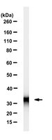
Abundance and subcellular localisation of cyclin D3 in human tumours.
Bartkova, J; Zemanova, M; Bartek, J
Int J Cancer
65
323-7
1996
Mostra il sommario
The D-type cyclins are positive regulators of the G1 phase of the mammalian cell cycle. Cyclins D1 or D2 are over-expressed in several types of cancer, transform rodent cells in culture and therefore harbor hallmarks of cellular proto-oncogenes. In contrast, no data on expression of cyclin D3 in tissues and tumours are presently available. We have raised monoclonal antibodies (MAbs) specific for cyclin D3 and examined abundance and subcellular localisation of this G1 cyclin in a series of human cultured cell types and in 180 primary tumours of diverse histogenesis. Cyclin D3 localised predominantly in nuclei of normal and tumour cells both in culture and in situ, and a pronounced cell-to-cell variation of its abundance was reminiscent of cyclins D1 and D2. Immunohistochemical analysis of tumour and corresponding normal tissues showed strong aberrant accumulation of cyclin D3 in a subset (about 10%) of breast carcinomas, whereas only weak-to-moderate expression was found in colorectal, head and neck and uterine carcinomas, melanomas and soft tissue sarcomas. The specificity of the immunohistochemical data was confirmed by immunoblotting analysis of tissue and tumour lysates. Our results indicate that over-abundance of cyclin D3 is considerably less frequent than that of cyclin D1, yet we identify subsets of breast tumours, and potentially lymphomas, as candidate tumour types with elevated cyclin D3 expression. | 8575852
|