The pathogenic role of the canonical Wnt pathway in age-related macular degeneration.
Zhou, T; Hu, Y; Chen, Y; Zhou, KK; Zhang, B; Gao, G; Ma, JX
Investigative ophthalmology & visual science
51
4371-9
2009
Mostra il sommario
The authors' previous studies showed that the Wnt signaling pathway is activated in the retinas and retinal pigment epithelia of animal models of age-related macular degeneration (AMD) and diabetic retinopathy (DR). The purpose of this study was to investigate the role of the canonical Wnt pathway in pathogenesis of these diseases.The Wnt pathway was activated using the Wnt3a-conditioned medium and adenovirus expressing a constitutively active mutant of beta-catenin (Ad-S37A) in ARPE19, a cell line derived from human RPE. Ad-S37A was injected into the vitreous of normal rats to activate the Wnt pathway in the retina. Accumulation of beta-catenin was determined by Western blot analysis, and its nuclear translocation was revealed by immunocytochemistry. Inflammatory factors were quantified by Western blot analysis and ELISA. Oxidative stress was determined by measuring intracellular reactive oxygen species (ROS) generation and nitrotyrosine levels.The Wnt3a-conditioned medium and Ad-S37A both increased beta-catenin levels and its nuclear translocation in ARPE19 cells, suggesting activation of the canonical Wnt pathway. Activation of the Wnt pathway significantly upregulated the expression of VEGF, NF-kappaB, and TNF-alpha. Further, Ad-S37A induced ROS generation in a dose-dependent manner. Wnt3a also induced a twofold increase of ROS generation. Intravitreal injection of Ad-S37A upregulated the expression of VEGF, ICAM-1, NF-kappaB, and TNF-alpha and increased protein nitrotyrosine levels in the retinas of normal rats.Activation of the canonical Wnt pathway is sufficient to induce retinal inflammation and oxidative stress and plays a pathogenic role in AMD and DR. Testo completo dell'articolo | 19875668
 |
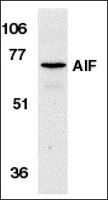
Molecular characterization of mitochondrial apoptosis-inducing factor.
Susin, S A, et al.
Nature, 397: 441-6 (1999)
1998
Mostra il sommario
Mitochondria play a key part in the regulation of apoptosis (cell death). Their intermembrane space contains several proteins that are liberated through the outer membrane in order to participate in the degradation phase of apoptosis. Here we report the identification and cloning of an apoptosis-inducing factor, AIF, which is sufficient to induce apoptosis of isolated nuclei. AIF is a flavoprotein of relative molecular mass 57,000 which shares homology with the bacterial oxidoreductases; it is normally confined to mitochondria but translocates to the nucleus when apoptosis is induced. Recombinant AIF causes chromatin condensation in isolated nuclei and large-scale fragmentation of DNA. It induces purified mitochondria to release the apoptogenic proteins cytochrome c and caspase-9. Microinjection of AIF into the cytoplasm of intact cells induces condensation of chromatin, dissipation of the mitochondrial transmembrane potential, and exposure of phosphatidylserine in the plasma membrane. None of these effects is prevented by the wide-ranging caspase inhibitor known as Z-VAD.fmk. Overexpression of Bcl-2, which controls the opening of mitochondrial permeability transition pores, prevents the release of AIF from the mitochondrion but does not affect its apoptogenic activity. These results indicate that AIF is a mitochondrial effector of apoptotic cell death. | 9989411
 |


