Millipore Sigma Vibrant Logo

ABE605 Anti-TREX1 Antibody

View Products on Sigmaaldrich.com
ABE605
100 µg  
Prix en cours de récupération
Le prix n'a pas pu être récupéré
La quantité minimale doit être un multiple de
Maximum Quantity is
À la validation de la commande Plus d'informations
Vous avez sauvegardé ()
 
Demander le prix
Disponibilité limitée
En stock 
Interrompu(e)
Disponible en quantités limitées
Disponibilité à confirmer
    Pour le restant : Nous vous tiendrons informé
      Pour le restant : Nous vous tiendrons informé
      Nous vous tiendrons informé
      Contacter le Service Clients
      Contact Customer Service

      Offres spéciales


      Contacter le Service Clients

      Aperçu

      Replacement Information

      Offres spéciales

      Tableau de caractéristiques principal

      Species ReactivityKey ApplicationsHostFormatAntibody Type
      HWB, IHC, IFRbAffinity PurifiedPolyclonal Antibody
      Description
      Catalogue NumberABE605
      DescriptionAnti-TREX1 Antibody
      Alternate Names
      • Three prime repair exonuclease 1
      • CRV
      • AGS1
      • DRN3
      • HERNS
      • 3'-5' exonuclease TREX1
      • DNase III
      Background InformationTREX1, (DNase III), or three prime repair exonuclease 1 is the major 3’→ 5’ DNA exonuclease in mammalian cells. TREX1 binds preferentially to ssDNA and can also remove mismatched 3’ terminal deoxyribonucleotides at DNA strand breaks. The protein is required for proper DNA checkpoint signaling and plays critical roles in DNA replication and gap filling during DNA repair. TREX1 has a perinuclear distribution in mammalian cells and is colocalized with ER-associated proteins in cells at rest. TREX1 relocalizes to the nucleus and specifically to replication forks during S phase and periods of DNA synthesis and repair such as after irradiation. TREX1 acts on an ssDNA polynucleotide species arising from the processing of aberrant DNA replication intermediates and thus acts to attenuate DNA damage checkpoint signaling and prevent pathological immune activation in normal cells. Defects in TREX1 protein have been associated in humans with many inherited diseases including Aicardi-Goutiėres syndrome type 1 (AGS1), systemic lupus erythematosus (SLE), chilblain lupus type 1 (CHBL1), and vasculopathy, retinal, with cerebral leukodystrophy (RVCL). EMD Millipore’s Anti-TREX1 rabbit polyclonal antibody has been tested in western blotting, immunohistochemistry, and immunofluorescence in human samples.
      References
      Product Information
      FormatAffinity Purified
      Control
      • Human spleen tissue lysate
      PresentationPurified rabbit polyclonal in buffer containing PBS with up to 0.1% sodium azide.
      Quality LevelMQ100
      Applications
      ApplicationDetect DNASE using this rabbit polyclonal antibody, Anti-TREX1 Antibody validated for use in western blotting, IHC & Immunofluorescence.
      Key Applications
      • Western Blotting
      • Immunohistochemistry
      • Immunofluorescence
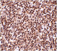
      Application NotesImmunohistochemistry Analysis: 2.5 µg/mL from a representative lot detected TREX1 in human spleen tissue.

      Immunofluorescence Analysis: 20 μg/mL from a representative lot detected TREX1 in human spleen tissue.
      Biological Information
      ImmunogenKLH-conjugated linear peptide corresponding to human TREX1.
      ConcentrationPlease refer to the Certificate of Analysis for the lot-specific concentration.
      HostRabbit
      Species Reactivity
      • Human
      Antibody TypePolyclonal Antibody
      Gene Symbol
      • TREX1
      Purification MethodAntigen Affinity Purified
      UniProt Number
      Molecular Weight~30 kDa observed. Uncharacterized bands may appear in some lysate(s). Uniprot describes 3 isoforms at ~32 kDa, ~33 kDa, and ~39 kDa.
      Physicochemical Information
      Dimensions
      Materials Information
      Toxicological Information
      Safety Information according to GHS
      Safety Information
      Product Usage Statements
      Quality AssuranceEvaluated by Western Blotting in human spleen tissue lysate.

      Western Blotting Analysis: 1 µg/mL of this antibody detected TREX1 in 15 µg of human spleen tissue lysate.
      Usage Statement
      • Unless otherwise stated in our catalog or other company documentation accompanying the product(s), our products are intended for research use only and are not to be used for any other purpose, which includes but is not limited to, unauthorized commercial uses, in vitro diagnostic uses, ex vivo or in vivo therapeutic uses or any type of consumption or application to humans or animals.
      Storage and Shipping Information
      Storage ConditionsStable for 1 year at 2-8°C from date of receipt.
      Packaging Information
      Material Size100 µg
      Transport Information
      Supplemental Information
      Specifications
      Global Trade Item Number
      Référence GTIN
      ABE605 04053252905469