Millipore Sigma Vibrant Logo

MABC966 Anti-Perforin-1 Antibody, clone dG9

MABC966
100 μg  
Prix en cours de récupération
Le prix n'a pas pu être récupéré
La quantité minimale doit être un multiple de
Maximum Quantity is
À la validation de la commande Plus d'informations
Vous avez sauvegardé ()
 
Demander le prix
Disponibilité limitée
En stock 
Interrompu(e)
Disponible en quantités limitées
Disponibilité à confirmer
    Pour le restant : Nous vous tiendrons informé
      Pour le restant : Nous vous tiendrons informé
      Nous vous tiendrons informé
      Contacter le Service Clients
      Contact Customer Service

      Offres spéciales


      Contacter le Service Clients

      Aperçu

      Replacement Information

      Offres spéciales

      Tableau de caractéristiques principal

      Species ReactivityKey ApplicationsHostFormatAntibody Type
      HFC, IP, ICC, IHCMPurifiedMonoclonal Antibody
      Description
      Catalogue NumberMABC966
      DescriptionAnti-Perforin-1 Antibody, clone dG9
      Alternate Names
      • Perforin-1
      • P1
      • Cytolysin
      • Lymphocyte pore-forming protein
      • PFP
      • Clone deltaG9
      Background InformationPerforin-1 (UniProt P14222; also known as Cytolysin, Lymphocyte pore forming protein, P1, PFP) is encoded by the PRF1 (Also known as FLH2, HPLH2, PFN1, PFP) gene in human (Gene ID 5551). Perforin is a secreted protein synthesized by activated cytotoxic T lymphocytes (CTL) and natural killer (NK) cells. It is a key component of the secreted lytic granule of CTL and NK cells. Perforin inserts into the phospholipid bilayer of the target cell plasma membrane, and polymerises to form a pore of ∼16 nm that causes target cell death by osmotic lysis and by allowing other lytic granule components, most notably granzyme B, to enter the targeted cells. Perforin is synthesized as a 70 kDa inactive precursor which is cleaved at the C-terminus to yield a 60 kDa active form.
      References
      Product Information
      FormatPurified
      PresentationPurified mouse monoclonal IgG2bκ antibody in buffer containing 0.1 M Tris-Glycine (pH 7.4), 150 mM NaCl with 0.05% sodium azide.
      Quality LevelMQ300
      Applications
      ApplicationAnti-Perforin-1 Antibody, clone dG9 is an antibody against Perforin-1 for use in Flow Cytometry, Immunoprecipitation, Immunocytochemistry, Immunohistochemistry.
      Key Applications
      • Flow Cytometry
      • Immunoprecipitation
      • Immunocytochemistry
      • Immunohistochemistry
      Application NotesImmunoprecipitation Analysis: A representative lot immunoprecipitated Perforin-1 in YT cell lysate with or without Endo H treatment (Uellner, R., et al. (1997). EMBO. 16(24):7287-7296).
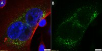
      Immunocytochemistry Analysis: A representative lot detected Perforin-1 in human lymphokine-activated killer (LAK) cells (Hameed, A., et al. (1992). American J. of Pathology. 140(5):1025-1030).
      Immunohistochemistry Analysis: A representative lot detected Perforin-1 in human cardiac biopsy samples during allograft rejection (Hameed, A., et al. (1992). American J. of Pathology. 140(5):1025-1030).
      Biological Information
      ImmunogenPurified granules from the human cytotoxic large granular lymphocytes lymphoma line YT corresponding to human Perforin-1.
      ClonedG9
      ConcentrationPlease refer to lot specific datasheet.
      HostMouse
      SpecificityReacts with non-denatured human perforin precursor and mature perforin, but not denatured perforin.
      IsotypeIgG2bκ
      Species Reactivity
      • Human
      Antibody TypeMonoclonal Antibody
      Entrez Gene Number
      Gene Symbol
      • PRF1
      • PFP
      Purification MethodProtein G Purified
      UniProt Number
      Molecular Weight61 kDa calculated
      Physicochemical Information
      Dimensions
      Materials Information
      Toxicological Information
      Safety Information according to GHS
      Safety Information
      Product Usage Statements
      Quality AssuranceEvaluated by Flow Cytometry in human peripheral blood mononuclear cells.

      Flow Cytometry Analysis: 3.0 µg of this antibody detected Perforin-1 in human peripheral blood mononuclear cells.
      Usage Statement
      • Unless otherwise stated in our catalog or other company documentation accompanying the product(s), our products are intended for research use only and are not to be used for any other purpose, which includes but is not limited to, unauthorized commercial uses, in vitro diagnostic uses, ex vivo or in vivo therapeutic uses or any type of consumption or application to humans or animals.
      Storage and Shipping Information
      Storage ConditionsStable for 1 year at 2-8°C from date of receipt.
      Packaging Information
      Material Size100 μg
      Transport Information
      Supplemental Information
      Specifications
      Global Trade Item Number
      Référence GTIN
      MABC966 04055977169843

      Documentation

      Anti-Perforin-1 Antibody, clone dG9 FDS

      Titre

      Fiche de données de sécurité des matériaux (FDS) 

      Anti-Perforin-1 Antibody, clone dG9 Certificats d'analyse

      TitreNuméro de lot
      Anti-Perforin-1, clone dG9 - 3590147 3590147
      Anti-Perforin-1, clone dG9 - 4083060 4083060
      Anti-Perforin-1, clone dG9 -Q2569117 Q2569117

      Informations techniques

      Titre
      Characterization of Estrogen Receptor α Phosphorylation Sites in Breast Cancer Tissue Using the SNAP i.d® 2.0 System
      White Paper: Further considerations of antibody validation and usage.