Pigment epithelium-derived factor (PEDF) protects cortical neurons in vitro from oxidant injury by activation of extracellular signal-regulated kinase (ERK) 1/2 and induction of Bcl-2.
Sanchez, A; Tripathy, D; Yin, X; Luo, J; Martinez, J; Grammas, P
Neuroscience research
72
1-8
2011
Afficher le résumé
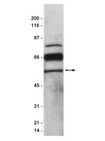
Mitigating oxidative stress-induced damage is critical to preserve neuronal function in diseased or injured brains. This study explores the mechanisms contributing to the neuroprotective effects of pigment epithelium-derived factor (PEDF) in cortical neurons. Cultured primary neurons are exposed to PEDF and H₂O₂ as well as inhibitors of phosphoinositide-3 kinase (PI3K) or extracellular signal-regulated kinase 1/2 (ERK1/2). Neuronal survival, cell death and levels of caspase 3, PEDF, phosphorylated ERK1/2, and Bcl-2 are measured. The data show cortical cultures release PEDF and that H₂O₂ treatment causes cell death, increases activated caspase 3 levels and decreases release of PEDF. Exogenous PEDF induces a dose-dependent increase in Bcl-2 expression and neuronal survival. Blocking Bcl-2 expression by siRNA reduced PEDF-induced increases in neuronal survival. Treating cortical cultures with PEDF 24 h before H₂O₂ exposure mitigates oxidant-induced decreases in neuronal survival, Bcl-2 expression, and phosphorylation of ERK1/2 and also reduces elevated caspase 3 level and activity. PEDF pretreatment effect on survival is blocked by inhibiting ERK or PI3K. However, only inhibition of ERK reduced the ability of PEDF to protect neurons from H₂O₂-induced Bcl-2 decrease and neuronal death. These data demonstrate PEDF-mediated neuroprotection against oxidant injury is largely mediated via ERK1/2 and Bcl-2 and suggest the utility of PEDF in preserving the viability of oxidatively challenged neurons. | Western Blotting | 21946416
 |
Multiple neurotrophic effects of VEGF on cultured neurons.
Sanchez, A; Wadhwani, S; Grammas, P
Neuropeptides
44
323-31
2009
Afficher le résumé
A large literature demonstrates the multifunctional nature of vascular endothelial growth factor (VEGF). Though initially characterized as an endothelial cell-specific factor, recent studies reveal that VEGF has numerous effects on diverse cell types in the brain including neurons. The objective of this study is to examine the effects of VEGF in cultured cortical neurons on survival, p38 mitogen-activated protein kinase (p38 MAP kinase) activity, pro- and anti-apoptotic protein expression and on release of neurotrophic and neurotoxic factors. The results show that VEGF dose-dependently enhances the survival of neurons in culture. VEGF decreases active caspase 3 levels and increases expression of the anti-apoptotic protein Bcl-2. VEGF decreases phosphorylated p38 MAP kinase level and activity in cortical neurons. In addition to modulating survival/death pathways in cortical neurons, VEGF also regulates release of proteins that affect neuronal viability. VEGF causes a dose-dependent release of the neurotrophic protein pigment epithelial-derived factor (PEDF), while significantly decreasing release of the neurotoxic protein amyloid beta. The VEGF-mediated decrease in amyloid beta is dependent on a functional Flt-1 receptor and is inhibited by dicoumarol, a multifunctional inhibitor of stress-activated protein kinase (SAPK)/JNK and NFkappaB pathways. Taken together, these data demonstrate that the neurotrophic effects of VEGF are likely mediated directly by increasing survival and decreasing apoptotic proteins and signals as well as indirectly by modulating release of proteins that affect neuronal viability. Article en texte intégral | | 20430442
 |
Localization of pigment epithelium derived factor (PEDF) in developing and adult human ocular tissues.
Karakousis, P C, et al.
Mol. Vis., 7: 154-63 (2001)
2001
Afficher le résumé
PURPOSE: To localize pigment epithelium-derived factor (PEDF) in developing and adult human ocular tissues. METHODS: PEDF was localized in fetal and adult eyes by immunofluorescence with a polyclonal antibody (pAb) against amino acids 327-343 of PEDF, or a monoclonal antibody (mAb) against the C-terminal 155 amino acids of PEDF. Specificity of the antibodies was documented by Western blotting. PEDF mRNA was localized in adult retina by in situ hybridization. RESULTS: In developing retinas (7.4 to 21.5 fetal weeks, Fwks), pAb anti-PEDF labeled retinal pigment epithelium (RPE) granules, developing cones, some neuroblasts and many cells in the ganglion cell layer (GCL). In adult retinas, pAb anti-PEDF labeled rod and cone cytoplasm and nuclei of rods but not cones. Cells in the INL and GCL, choroid, corneal epithelium and endothelium, and ciliary body were also pAb PEDF-positive. Preadsorption of pAb anti-PEDF with the immunizing peptide blocked specific labeling in retina and other tissues, except for photoreceptor outer segments. In agreement with the immunolocalization with pAb anti-PEDF, in situ hybridization revealed PEDF mRNA in the RPE, photoreceptors, inner nuclear layer cells and ganglion cells in adult retina. In developing retinas 18 Fwks and older, and in adult retinas, mAb anti-PEDF labeled the interphotoreceptor matrix (IPM). Western blots of retina, cornea, and ciliary body/iris with pAb anti-PEDF produced several bands at about 46 kDa. With mAb anti-PEDF, retina produced one band at about 46 kDa; cornea and ciliary body/iris had several bands at about 46 kDa. CONCLUSIONS: PEDF, originally reported as a product of RPE cells, is present in photoreceptors and inner retinal cell types in developing and adult human eyes. Photoreceptors and RPE may secrete PEDF into the IPM. | | 11438800
 |


