Millipore Sigma Vibrant Logo

MABS1954-100UG Anti-DARC (CD234) Antibody, clone NaM185-2C3

View Products on Sigmaaldrich.com
MABS1954-100UG
100 μg  
Prix en cours de récupération
Le prix n'a pas pu être récupéré
La quantité minimale doit être un multiple de
Maximum Quantity is
À la validation de la commande Plus d'informations
Vous avez sauvegardé ()
 
Demander le prix
Disponibilité limitée
En stock 
Interrompu(e)
Disponible en quantités limitées
Disponibilité à confirmer
    Pour le restant : Nous vous tiendrons informé
      Pour le restant : Nous vous tiendrons informé
      Nous vous tiendrons informé
      Contacter le Service Clients
      Contact Customer Service


      Contacter le Service Clients

      Aperçu

      Replacement Information
      Description
      Catalogue NumberMABS1954-100UG
      DescriptionAnti-DARC (CD234) Antibody, clone NaM185-2C3
      Alternate Names
      • Atypical chemokine receptor 1
      • Duffy antigen/chemokine receptor
      • Fy glycoprotein
      • GpFy
      • Glycoprotein D
      • Plasmodium vivax receptor
      • CD234
      • Fy6
      Background InformationAtypical chemokine receptor 1 (UniProt: Q16570; also known as Duffy antigen/chemokine receptor, DARC, Fy glycoprotein, GpFy, Glycoprotein D, Plasmodium vivax receptor, CD234) is encoded by the ACKR1 (also known as DARC, FY, GPD) gene (Gene ID: 2532) in human. DARC is a multi-pass membrane protein of the G-protein coupled receptor 1 family with four extracellular domains, seven transmembrane domains, and four cytoplasmic domains. Its N-terminal glycosylated extracellular domain carries the Duffy blood group antigens Fya and Fyb. It is present mainly on erythrocytes and endothelial cells of post-capillary venules of various tissues. DARC serves as a promiscuous receptor for a number of pro-inflammatory CC and CXC chemokines but lacks signal transduction ability. However, it controls chemokine levels and localization via high-affinity chemokine binding that is uncoupled from classic ligand-driven signal transduction cascades, resulting instead in chemokine sequestration, degradation, or transcytosis. It is reported to regulate chemokine bioavailability and, consequently, leukocyte recruitment through two distinct mechanisms. When expressed in endothelial cells, it sustains the abluminal to luminal transcytosis of tissue-derived chemokines and their subsequent presentation to circulating leukocytes. However, when expressed in erythrocytes, it serves as blood reservoir of cognate chemokines and a chemokine sink, buffering potential surges in plasma chemokine levels. Clone NaM185-2C3 recognizes a linear epitope, the essential portion of which is localized in amino acids 22-26 where all amino acid residues of the epitope, except Asp, are essential for antibody-binding. (Ref.: Grodecka, M., et al. (2010). Acta Biochimica Polonica. 57(1); 49-53; Wasniowska, K., et al. (2002). Transfusion Med. 12(3); 205-211).
      References
      Product Information
      FormatPurified
      PresentationPurified mouse monoclonal antibody IgG1 in PBS without azide.
      Quality LevelMQ200
      Applications
      ApplicationAnti-DARC (CD234), clone NaM185-2C3, Cat. No. MABS1954, is a mouse monoclonal antibody that detects DARC (Duffy antigen/chemokine receptor) and is tested for use in ELISA, Flow Cytometry, Inhibition Assay, Radioimmunoassay, and Western Blotting.
      Key Applications
      • Flow Cytometry
      • ELISA
      • Radioimmunoassay
      • Western Blotting
      • Inhibition
      Application NotesRadioimmunoassay: A representative lot detected DARC (CD234) in Radioimmunoassay applications (Wasniowska, K., et. al. (2002). Transfus Med. 12(3):205-11).

      Western Blotting Analysis: A representative lot detected DARC (CD234) in Western Blotting applications (Grodecka, M., et. al. (2010). Acta Biochim Pol. 57(1):49-53).

      Flow Cytometry Analysis: A representative lot detected DARC (CD234) in Flow Cytometry applications (Menard, D., et. al. (2010). Proc Natl Acad Sci USA. 107(13):5967-71; Chi, J.S., et. al. (2016). Int J Parasitol. 46(1):31-9).

      ELISA Analysis: A representative lot detected DARC (CD234) in ELISA applications (Wasniowska, K., et. al. (2002). Transfus Med. 12(3):205-11).

      Inhibition Assay: A representative lot of this antibody blocked CXCL8 binding site on endothelial cells. (Whittall, C., et al. (2013). J. Immunol. 190(4):1725-36).

      Note: Actual optimal working dilutions must be determined by end user as specimens, and experimental conditions may vary with the end user
      Biological Information
      ImmunogenCHO cells transfected with cDNA encoding full-length Duffy antigen/chemokine receptor (DARC) protein.
      EpitopeN-terminal
      CloneNaM185-2C3
      Concentration1.0 mg/mL. Please refer to guidance on suggested starting dilutions and/or titers per application and sample type.
      HostMouse
      SpecificityClone NaM185-2C3 (2C3) is a mouse monoclonal antibody that detects DARC (CD234). It targets an epitope within 8 amino acids from the N-terminal region.
      IsotypeIgG1κ
      Species Reactivity
      • Human
      Species Reactivity NoteHuman.
      Antibody TypeMonoclonal Antibody
      Entrez Gene Number
      Gene Symbol
      • ACKR1
      • DARC
      • FT
      • GPD
      Purification MethodProtein G purified
      UniProt Number
      Molecular Weight35.55 kDa calculated.
      Physicochemical Information
      Dimensions
      Materials Information
      Toxicological Information
      Safety Information according to GHS
      Safety Information
      Product Usage Statements
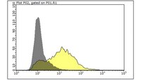
      Quality AssuranceEvaluated by Flow Cytometry in K562 cells.

      Flow Cytometry Analysis: 1 µg of this antibody detected DARC (CD234) in one million K562 cells.
      Usage Statement
      • Unless otherwise stated in our catalog or other company documentation accompanying the product(s), our products are intended for research use only and are not to be used for any other purpose, which includes but is not limited to, unauthorized commercial uses, in vitro diagnostic uses, ex vivo or in vivo therapeutic uses or any type of consumption or application to humans or animals.
      Storage and Shipping Information
      Storage ConditionsStable for 1 year at -10°C to -25°C from date of receipt. Handling Recommendations: Upon receipt and prior to removing the cap, centrifuge the vial and gently mix the solution. Aliquot into microcentrifuge tubes and store at -20°C. Avoid repeated freeze/thaw cycles, which may damage IgG and affect product performance.
      Packaging Information
      Material Size100 μg
      Transport Information
      Supplemental Information
      Specifications
      Global Trade Item Number
      Référence GTIN
      MABS1954-100UG 04065266020625