Our broad portfolio consists of multiplex panels that allow you to choose, within the panel, analytes that best meet your needs. On a separate tab you can choose the premixed cytokine format or a single plex kit.
Cell Signaling Kits & MAPmates™
Choose fixed kits that allow you to explore entire pathways or processes. Or design your own kits by choosing single plex MAPmates™, following the provided guidelines.
The following MAPmates™ should not be plexed together:
-MAPmates™ that require a different assay buffer
-Phospho-specific and total MAPmate™ pairs, e.g. total GSK3β and GSK3β (Ser 9)
-PanTyr and site-specific MAPmates™, e.g. Phospho-EGF Receptor and phospho-STAT1 (Tyr701)
-More than 1 phospho-MAPmate™ for a single target (Akt, STAT3)
-GAPDH and β-Tubulin cannot be plexed with kits or MAPmates™ containing panTyr
.
Catalogue Number
Ordering Description
Qty/Pack
List
This item has been added to favorites.
Select A Species, Panel Type, Kit or Sample Type
To begin designing your MILLIPLEX® MAP kit select a species, a panel type or kit of interest.
Custom Premix Selecting "Custom Premix" option means that all of the beads you have chosen will be premixed in manufacturing before the kit is sent to you.
Catalogue Number
Ordering Description
Qty/Pack
List
This item has been added to favorites.
Species
Panel Type
Selected Kit
Qty
Catalogue Number
Ordering Description
Qty/Pack
List Price
96-Well Plate
Qty
Catalogue Number
Ordering Description
Qty/Pack
List Price
Add Additional Reagents (Buffer and Detection Kit is required for use with MAPmates)
Qty
Catalogue Number
Ordering Description
Qty/Pack
List Price
48-602MAG
Buffer Detection Kit for Magnetic Beads
1 Kit
Space Saver Option Customers purchasing multiple kits may choose to save storage space by eliminating the kit packaging and receiving their multiplex assay components in plastic bags for more compact storage.
This item has been added to favorites.
The Product Has Been Added To Your Cart
You can now customize another kit, choose a premixed kit, check out or close the ordering tool.
NA81
Sigma-AldrichAnti-5-Methylcytosine Mouse mAb (162 33 D3)
This Anti-5-Methylcytosine Mouse mAb (162 33 D3) is validated for use in ELISA, FC, Frozen Sections, IF, Paraffin Sections, Radioimmunoassay, Southwestern Blot for the detection of 5-Methylcytosine.
More>>This Anti-5-Methylcytosine Mouse mAb (162 33 D3) is validated for use in ELISA, FC, Frozen Sections, IF, Paraffin Sections, Radioimmunoassay, Southwestern Blot for the detection of 5-Methylcytosine. Less<<
Anti-5-Methylcytosine Mouse mAb (162 33 D3) MSDS (material safety data sheet) or SDS, CoA and CoQ, dossiers, brochures and other available documents.
The quantity field is empty. Please enter a quantity of 1 or more to add items to your cart.
Description
Overview
Recognizes 5-methylcytosine in methylated DNA or RNA in NIH3T3 cells.
Specific • Affinity purified mouse monoclonal antibody - Detects methylated DNA from a broad range of species.
Several Applications • Flow Cytometry
• Frozen Sections
• Immunoblotting
• Immunofluorescence
• Paraffin Sections
Reliable • Generates reproducible results.
• Clone 162 33 D3 has been used in over 50 publications.
Catalogue Number
NA81
Brand Family
Calbiochem®
Synonyms
Anti-5-mc, Anti-5-MeCyd
Application Data
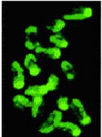
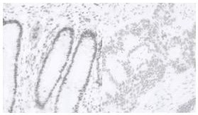
Detection of 5-methylcytosine by immunohistochemistry. Sample: Normal colon (left) and colon adenocarcinoma (right). Anti-5-Methylcytosine Mouse mAb (162 33 D3) (Cat. No. NA81). Detection: DAB Chromosome methylation patterns of mouse embryos at the one-cell stage. Methylated sites were revealed by indirect immunofluorescence labeling with Anti-5-methylcytosine.
References
References
Ehrlich, M. 2002. Oncogene21, 5400. (Review) Widschwendter, M. and Jones, P.A. 2002. Oncogene21, 5462. (Review) Barton, S.C., et al. 2001. Hum. Mol. Genet.10, 2983. Piyathilake, C.J., et al. 2001. Hum. Pathol.32, 856. Taddei, A., et al. 2001. Nat. Cell Biol.3, 114. Piyathilake, C.J., et al. 2000. Biotech. Histochem.75, 251. De Capoa, A., et al. 1999. FASEB J.13, 89.
Clone 162 33 D3 is useful for the quantitative and qualitative detection of methylated DNA or RNA in a variety of samples and applications. It has been used in over 40 publications. Antibody should be titrated for optimal results in an individual systems.
Biological Information
Immunogen
5-methylcytosine conjugated to ovalbumin
Clone
162 33 D3
Host
Mouse
Isotype
IgG₁
Species Reactivity
A Broad Range Of Species
Antibody Type
Monoclonal Antibody
Concentration Label
Please refer to vial label for lot-specific concentration
Storage and Shipping Information
Ship Code
Blue Ice Only
Toxicity
Standard Handling
Storage
-20°C
Avoid freeze/thaw
Avoid freeze/thaw
Do not freeze
Ok to freeze
Special Instructions
Following initial thaw, aliquot and freeze (-20°C).
Note that this data sheet is not lot-specific and is representative of the current specifications for this product. Please consult the vial label and the certificate of analysis for information on specific lots. Also note that shipping conditions may differ from storage conditions.
Detection of 5-methylcytosine by immunohistochemistry. Sample: Normal colon (left) and colon adenocarcinoma (right). Anti-5-Methylcytosine Mouse mAb (162 33 D3) (Cat. No. NA81). Detection: DAB Chromosome methylation patterns of mouse embryos at the one-cell stage. Methylated sites were revealed by indirect immunofluorescence labeling with Anti-5-methylcytosine.
DNA methylation plays an important role in gene regulation. Methylation of gene-promoter regions leads to loss of function while DNA demethylation can lead to gain of function. Alterations in DNA methylation may play an important role in carcinogenesis. DNA methylation varies according to tissue type. In some cancers types such as Wilms tumor and colon adenocarcinoma have a decrease in global DNA methylation. In contrast other cancers such as breast cancer have DNA hypermethylation. Changes in global DNA methylation status may be useful early markers for the detection of premalignant lesions.
Host
Mouse
Immunogen
5-methylcytosine conjugated to ovalbumin
Clone
162 33 D3
Isotype
IgG₁
Species
a broad range of species
Positive control
NIH3T3 cells
Form
Liquid
Formulation
In PBS, pH 7.4.
Concentration Label
Please refer to vial label for lot-specific concentration
Preservative
None
Comments
Clone 162 33 D3 is useful for the quantitative and qualitative detection of methylated DNA or RNA in a variety of samples and applications. It has been used in over 40 publications. Antibody should be titrated for optimal results in an individual systems.
Storage
Avoid freeze/thaw -20°C
Do Not Freeze
Ok to freeze
Special Instructions
Following initial thaw, aliquot and freeze (-20°C).
Toxicity
Standard Handling
References
Ehrlich, M. 2002. Oncogene21, 5400. (Review) Widschwendter, M. and Jones, P.A. 2002. Oncogene21, 5462. (Review) Barton, S.C., et al. 2001. Hum. Mol. Genet.10, 2983. Piyathilake, C.J., et al. 2001. Hum. Pathol.32, 856. Taddei, A., et al. 2001. Nat. Cell Biol.3, 114. Piyathilake, C.J., et al. 2000. Biotech. Histochem.75, 251. De Capoa, A., et al. 1999. FASEB J.13, 89.
Application references
Paraffin Sections
Kang, J.S., et al. 2006. Cancer Sci.97, 453.