Wenn Sie das Fenster schließen, wird Ihre Konfiguration nicht gespeichert, es sei denn, Sie haben Ihren Artikel in die Bestellung aufgenommen oder zu Ihren Favoriten hinzugefügt.
Klicken Sie auf OK, um das MILLIPLEX® MAP-Tool zu schließen oder auf Abbrechen, um zu Ihrer Auswahl zurückzukehren.
Wählen Sie konfigurierbare Panels & Premixed-Kits - ODER - Kits für die zelluläre Signaltransduktion & MAPmates™
Konfigurieren Sie Ihre MILLIPLEX® MAP-Kits und lassen sich den Preis anzeigen.
Konfigurierbare Panels & Premixed-Kits
Unser breites Angebot enthält Multiplex-Panels, für die Sie die Analyten auswählen können, die am besten für Ihre Anwendung geeignet sind. Unter einem separaten Register können Sie das Premixed-Cytokin-Format oder ein Singleplex-Kit wählen.
Kits für die zelluläre Signaltransduktion & MAPmates™
Wählen Sie gebrauchsfertige Kits zur Erforschung gesamter Signalwege oder Prozesse. Oder konfigurieren Sie Ihre eigenen Kits mit Singleplex MAPmates™.
Die folgenden MAPmates™ sollten nicht zusammen analysiert werden: -MAPmates™, die einen unterschiedlichen Assaypuffer erfordern. -Phosphospezifische und MAPmate™ Gesamtkombinationen wie Gesamt-GSK3β und Gesamt-GSK3β (Ser 9). -PanTyr und locusspezifische MAPmates™, z.B. Phospho-EGF-Rezeptor und Phospho-STAT1 (Tyr701). -Mehr als 1 Phospho-MAPmate™ für ein einziges Target (Akt, STAT3). -GAPDH und β-Tubulin können nicht mit Kits oder MAPmates™, die panTyr enthalten, analysiert werden.
.
Bestellnummer
Bestellinformationen
St./Pkg.
Liste
Dieser Artikel wurde zu Ihren Favoriten hinzugefügt.
Wählen Sie bitte Spezies, Panelart, Kit oder Probenart
Um Ihr MILLIPLEX® MAP-Kit zu konfigurieren, wählen Sie zunächst eine Spezies, eine Panelart und/oder ein Kit.
Custom Premix Selecting "Custom Premix" option means that all of the beads you have chosen will be premixed in manufacturing before the kit is sent to you.
Catalogue Number
Ordering Description
Qty/Pack
List
Dieser Artikel wurde zu Ihren Favoriten hinzugefügt.
Spezies
Panelart
Gewähltes Kit
Menge
Bestellnummer
Bestellinformationen
St./Pkg.
Listenpreis
96-Well Plate
Menge
Bestellnummer
Bestellinformationen
St./Pkg.
Listenpreis
Weitere Reagenzien hinzufügen (MAPmates erfordern die Verwendung eines Puffer- und Detektionskits)
Menge
Bestellnummer
Bestellinformationen
St./Pkg.
Listenpreis
48-602MAG
Buffer Detection Kit for Magnetic Beads
1 Kit
Platzsparende Option Kunden, die mehrere Kits kaufen, können ihre Multiplex-Assaykomponenten in Kunststoffbeuteln anstelle von Packungen erhalten, um eine kompaktere Lagerung zu ermöglichen.
Dieser Artikel wurde zu Ihren Favoriten hinzugefügt.
Das Produkt wurde in Ihre Bestellung aufgenommen
Sie können nun ein weiteres Kit konfigurieren, ein Premixed-Kit wählen, zur Kasse gehen oder das Bestell-Tool schließen.
LMR001
Sigma-AldrichCell Capture Imaging Reagent
Cell Capture Imaging Reagent is a specially formulated polymeric hydrogel that allows cells to be constrained at the bottom of microtiter plates for subsequent imaging.
More>>Cell Capture Imaging Reagent is a specially formulated polymeric hydrogel that allows cells to be constrained at the bottom of microtiter plates for subsequent imaging. Less<<
Empfohlene Produkte
Übersicht
Replacement Information
Description
Catalogue Number
LMR001
Description
Cell Capture Imaging Reagent
Alternate Names
Photopolymeric hydrogel
Background Information
Cell Capture Imaging Reagent is a specially formulated photopolymeric hydrogel thin film that is optically transparent and is permeable to antibodies and other immunocytochemistry reagents. It allows cells to be physically constrained at the bottom of standard imaging microtiter without any cell loss. It is particularly suitable for use in experiments where the cell population is less than 10,000. Cells captured in this hydrogel can be easily micrographed regardless of the staining reagents. This polymeric hydrogel displays sufficient mechanical strength to withstand routine pipetting and is dense enough for cells to sink to the imaging surface.
References
Product Information
Format
Liquid Reagent
Presentation
Clear viscous liquid.
Applications
Application
Cell Capture Imaging Reagent is a specially formulated polymeric hydrogel that allows cells to be constrained at the bottom of microtiter plates for subsequent imaging.
Key Applications
Immunocytochemistry
Application Notes
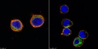
Immunocytochemistry Analysis: Use of this reagent with an antibody detected Jurkat cells.
1. Wash cells with 1x PBS twice 2. Re-suspend cells using PBS to concentration < 1 x 10⁶ cells / mL 3. In 384- Imaging well-plate, Add 40 µL of cell suspension per well. Number of cells will vary depending on user preference. 4. Add 6.5 µL of Cell Capture Imaging Reagent. Do not mix. Gently dispense using pipette.
NOTE: This ratio of cell volume to Cell Capture Imaging Reagent reagent volume is valid when using 384 Corning High Content microplate with glass bottom plates (Catalog Number 4581). If using a different plate with different dimension this ratio must be recalculated: Example: • 384 Corning High Content microplate with glass bottom plate (4581): .0625 cm2 • 96 A/2 Corning High Content microplate (4580): .1586 cm2 • Ratio: .1586/.0625= 2.54 Conversion Factor • Cells: 40ul*2.54= 101.5ul of Cells • Cell Capture Imaging Reagent: 6.5ul*2.54= 16.5 of Cell Capture Imaging Reagent
5. Centrifuge well-plate for 5 mins @ 500 g 6. Expose to UV-light for 15 seconds on ChemiDoc with standard UV wavelength, 302 nm NOTE: Exposure time may need to adjust based on UV intensity. UV light must come from the bottom of the well. To optimize UV exposure, a short time course with the correct ratio of PBS to Cell Capture Imaging Reagent can be used. To visualize formation, hold 96 well plate up to light to distinguish a slight yellow color and by taking a pipet tip and remove the gel plug from well. Use the shortest amount of UV exposure possible. Alternatively, to optimize UV exposure, take any cells resuspended in PBS with the appropriate amount of Cell Capture Imaging Reagent and expose to UV. Start with the shortest amount of time followed by steps 7-9 below and by a 10 min DAPI stain and washing with PBS. Visualize on microscope to make sure cell retention is high. This same procedure can also be used to determine a suitable number of cells to dispense into well. 7. Remove supernatant from each well by flicking plate 8. Add 1x PBS each well. Take caution not to poke the bottom layer. Discard supernatant by flicking the plate. 9. Repeat step 8 four times. 10. Cells are ready for immunocytochemistry. All immunocytochemistry steps can be performed in the well with Cell Capture Imaging Reagent encapsulated cells. Be sure to keep wells hydrated with PBS or buffer at all times.
Usage Statement
Unless otherwise stated in our catalog or other company documentation accompanying the product(s), our products are intended for research use only and are not to be used for any other purpose, which includes but is not limited to, unauthorized commercial uses, in vitro diagnostic uses, ex vivo or in vivo therapeutic uses or any type of consumption or application to humans or animals.