Wenn Sie das Fenster schließen, wird Ihre Konfiguration nicht gespeichert, es sei denn, Sie haben Ihren Artikel in die Bestellung aufgenommen oder zu Ihren Favoriten hinzugefügt.
Klicken Sie auf OK, um das MILLIPLEX® MAP-Tool zu schließen oder auf Abbrechen, um zu Ihrer Auswahl zurückzukehren.
Wählen Sie konfigurierbare Panels & Premixed-Kits - ODER - Kits für die zelluläre Signaltransduktion & MAPmates™
Konfigurieren Sie Ihre MILLIPLEX® MAP-Kits und lassen sich den Preis anzeigen.
Konfigurierbare Panels & Premixed-Kits
Unser breites Angebot enthält Multiplex-Panels, für die Sie die Analyten auswählen können, die am besten für Ihre Anwendung geeignet sind. Unter einem separaten Register können Sie das Premixed-Cytokin-Format oder ein Singleplex-Kit wählen.
Kits für die zelluläre Signaltransduktion & MAPmates™
Wählen Sie gebrauchsfertige Kits zur Erforschung gesamter Signalwege oder Prozesse. Oder konfigurieren Sie Ihre eigenen Kits mit Singleplex MAPmates™.
Die folgenden MAPmates™ sollten nicht zusammen analysiert werden: -MAPmates™, die einen unterschiedlichen Assaypuffer erfordern. -Phosphospezifische und MAPmate™ Gesamtkombinationen wie Gesamt-GSK3β und Gesamt-GSK3β (Ser 9). -PanTyr und locusspezifische MAPmates™, z.B. Phospho-EGF-Rezeptor und Phospho-STAT1 (Tyr701). -Mehr als 1 Phospho-MAPmate™ für ein einziges Target (Akt, STAT3). -GAPDH und β-Tubulin können nicht mit Kits oder MAPmates™, die panTyr enthalten, analysiert werden.
.
Bestellnummer
Bestellinformationen
St./Pkg.
Liste
Dieser Artikel wurde zu Ihren Favoriten hinzugefügt.
Wählen Sie bitte Spezies, Panelart, Kit oder Probenart
Um Ihr MILLIPLEX® MAP-Kit zu konfigurieren, wählen Sie zunächst eine Spezies, eine Panelart und/oder ein Kit.
Custom Premix Selecting "Custom Premix" option means that all of the beads you have chosen will be premixed in manufacturing before the kit is sent to you.
Catalogue Number
Ordering Description
Qty/Pack
List
Dieser Artikel wurde zu Ihren Favoriten hinzugefügt.
Spezies
Panelart
Gewähltes Kit
Menge
Bestellnummer
Bestellinformationen
St./Pkg.
Listenpreis
96-Well Plate
Menge
Bestellnummer
Bestellinformationen
St./Pkg.
Listenpreis
Weitere Reagenzien hinzufügen (MAPmates erfordern die Verwendung eines Puffer- und Detektionskits)
Menge
Bestellnummer
Bestellinformationen
St./Pkg.
Listenpreis
48-602MAG
Buffer Detection Kit for Magnetic Beads
1 Kit
Platzsparende Option Kunden, die mehrere Kits kaufen, können ihre Multiplex-Assaykomponenten in Kunststoffbeuteln anstelle von Packungen erhalten, um eine kompaktere Lagerung zu ermöglichen.
Dieser Artikel wurde zu Ihren Favoriten hinzugefügt.
Das Produkt wurde in Ihre Bestellung aufgenommen
Sie können nun ein weiteres Kit konfigurieren, ein Premixed-Kit wählen, zur Kasse gehen oder das Bestell-Tool schließen.
Anti-pan-ARF, clone 1D9, Cat. No. MABS2041, is a mouse monoclonal antibody that detects all ADP-ribosylation factors and has been tested for use in ELISA, Electron Microscopy, Immunoprecipitation, and Western Blotting,
More>>Anti-pan-ARF, clone 1D9, Cat. No. MABS2041, is a mouse monoclonal antibody that detects all ADP-ribosylation factors and has been tested for use in ELISA, Electron Microscopy, Immunoprecipitation, and Western Blotting, Less<<
Anti-pan-ARF Antibody, clone 1D9: SDB (Sicherheitsdatenblätter), Analysenzertifikate und Qualitätszertifikate, Dossiers, Broschüren und andere verfügbare Dokumente.
ADP-ribosylation factor 1 (UniProt: P84077; also known as Arf1) is encoded by the ARF1 gene (Gene ID: 375) in human. Arf1 is a member of the family of low molecular weight GTP-binding proteins. It is abundant in neural tissues where it may comprise up to 1% of total cellular protein. The ARF proteins are categorized as class I (ARF1, ARF2 and ARF3), class II (ARF4 and ARF5) and class III (ARF6), and members of each class share a common gene organization. Arf1, 3, 4, and 5 are predominantly cytosolic, but could be recruited to a variety of intracellular membranes, but not plasma membranes, upon incubation in the presence of GTP S. Arf6 is found at the plasma membrane and in endosomes. Arf1 is localized to the Golgi apparatus and has a central role in intra-Golgi transport. It has two nucleotide binding regions (aa 24-32 and 126-129). Arf1 functions as an allosteric activator of the cholera toxin catalytic subunit, an ADP-ribosyltransferase. It is also involved in protein trafficking among different compartments and is reported to modulate vesicle budding and uncoating within the Golgi complex. In its GTP-bound form, its triggers the association with coat proteins with the Golgi membrane. The hydrolysis of Arf1-bound GTP, which is mediated by ARFGAPs proteins, is required for dissociation of coat proteins from Golgi membranes and vesicles. This antibody (clone 1D9) detects all Arf proteins, but to different degrees. (Ref.: Cavenagh, MM., et al. (1996). J. Biol. Chem. 271(36):21767-74).
References
Product Information
Format
Purified
Presentation
Purified mouse monoclonal antibody IgG1 in buffer containing 0.1 M Tris-Glycine (pH 7.4), 150 mM NaCl with 0.05% sodium azide.
Anti-pan-ARF, clone 1D9, Cat. No. MABS2041, is a mouse monoclonal antibody that detects all ADP-ribosylation factors and has been tested for use in ELISA, Electron Microscopy, Immunoprecipitation, and Western Blotting,
Key Applications
ELISA
Electron Microscopy
Immunoprecipitation
Western Blotting
Application Notes
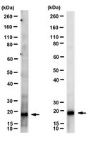
Western Blotting Analysis: 1 ug/mL from a representative lot detected ARF proteins in human lung tissue lysate.
Immunoprecipitation Analysis: A representative lot immunoprecipitated ARF proteins (Cavenagh, M.M., et. al. (1996). J Biol Chem. 271(36):21767-74).
ELISA Analysis: A representative lot detected pan-ARF in ELISA applications (Cavenagh, M.M., et. al. (1996). J Biol Chem. 271(36):21767-74).
Western Blotting Analysis: A representative lot detected ARF proteins in Western Blotting applications (Cavenagh, M.M., et. al. (1996). J Biol Chem. 271(36):21767-74; Zuezem, S., et. al. (1992). Proc Natl Acad Sci USA. 89(14):6619-23).
Electron Microscopy Analysis: A representative lot detected ARF proteins in Electron Microscopy applications (Zuezem, S., et. al. (1992). Proc Natl Acad Sci USA. 89(14):6619-23).
Dot Blot Analysis: A representative lot detected ARF proteins in Dot Blot applications (Cavenagh, M.M., et. al. (1996). J Biol Chem. 271(36):21767-74).
Biological Information
Immunogen
Purified full length human recombinant ADP-ribosylation factor 1.
Clone
1D9
Concentration
Please refer to lot specific datasheet.
Host
Mouse
Specificity
Clone 1D9 detects all isoforms of DP-ribosylation factors in human cells.
~20 kDa observed. Uncharacterized bands may be observed in some lysate(s).
Physicochemical Information
Dimensions
Materials Information
Toxicological Information
Safety Information according to GHS
Safety Information
Product Usage Statements
Quality Assurance
Evaluated by Western Blotting in HeLa cell lysate.
Western Blotting Analysis: 1 ug/mL of this antibody detected pan-ARF in HeLa cell lysate.
Usage Statement
Unless otherwise stated in our catalog or other company documentation accompanying the product(s), our products are intended for research use only and are not to be used for any other purpose, which includes but is not limited to, unauthorized commercial uses, in vitro diagnostic uses, ex vivo or in vivo therapeutic uses or any type of consumption or application to humans or animals.