Wenn Sie das Fenster schließen, wird Ihre Konfiguration nicht gespeichert, es sei denn, Sie haben Ihren Artikel in die Bestellung aufgenommen oder zu Ihren Favoriten hinzugefügt.
Klicken Sie auf OK, um das MILLIPLEX® MAP-Tool zu schließen oder auf Abbrechen, um zu Ihrer Auswahl zurückzukehren.
Wählen Sie konfigurierbare Panels & Premixed-Kits - ODER - Kits für die zelluläre Signaltransduktion & MAPmates™
Konfigurieren Sie Ihre MILLIPLEX® MAP-Kits und lassen sich den Preis anzeigen.
Konfigurierbare Panels & Premixed-Kits
Unser breites Angebot enthält Multiplex-Panels, für die Sie die Analyten auswählen können, die am besten für Ihre Anwendung geeignet sind. Unter einem separaten Register können Sie das Premixed-Cytokin-Format oder ein Singleplex-Kit wählen.
Kits für die zelluläre Signaltransduktion & MAPmates™
Wählen Sie gebrauchsfertige Kits zur Erforschung gesamter Signalwege oder Prozesse. Oder konfigurieren Sie Ihre eigenen Kits mit Singleplex MAPmates™.
Die folgenden MAPmates™ sollten nicht zusammen analysiert werden: -MAPmates™, die einen unterschiedlichen Assaypuffer erfordern. -Phosphospezifische und MAPmate™ Gesamtkombinationen wie Gesamt-GSK3β und Gesamt-GSK3β (Ser 9). -PanTyr und locusspezifische MAPmates™, z.B. Phospho-EGF-Rezeptor und Phospho-STAT1 (Tyr701). -Mehr als 1 Phospho-MAPmate™ für ein einziges Target (Akt, STAT3). -GAPDH und β-Tubulin können nicht mit Kits oder MAPmates™, die panTyr enthalten, analysiert werden.
.
Bestellnummer
Bestellinformationen
St./Pkg.
Liste
Dieser Artikel wurde zu Ihren Favoriten hinzugefügt.
Wählen Sie bitte Spezies, Panelart, Kit oder Probenart
Um Ihr MILLIPLEX® MAP-Kit zu konfigurieren, wählen Sie zunächst eine Spezies, eine Panelart und/oder ein Kit.
Custom Premix Selecting "Custom Premix" option means that all of the beads you have chosen will be premixed in manufacturing before the kit is sent to you.
Catalogue Number
Ordering Description
Qty/Pack
List
Dieser Artikel wurde zu Ihren Favoriten hinzugefügt.
Spezies
Panelart
Gewähltes Kit
Menge
Bestellnummer
Bestellinformationen
St./Pkg.
Listenpreis
96-Well Plate
Menge
Bestellnummer
Bestellinformationen
St./Pkg.
Listenpreis
Weitere Reagenzien hinzufügen (MAPmates erfordern die Verwendung eines Puffer- und Detektionskits)
Menge
Bestellnummer
Bestellinformationen
St./Pkg.
Listenpreis
48-602MAG
Buffer Detection Kit for Magnetic Beads
1 Kit
Platzsparende Option Kunden, die mehrere Kits kaufen, können ihre Multiplex-Assaykomponenten in Kunststoffbeuteln anstelle von Packungen erhalten, um eine kompaktere Lagerung zu ermöglichen.
Dieser Artikel wurde zu Ihren Favoriten hinzugefügt.
Das Produkt wurde in Ihre Bestellung aufgenommen
Sie können nun ein weiteres Kit konfigurieren, ein Premixed-Kit wählen, zur Kasse gehen oder das Bestell-Tool schließen.
Detect Thrombospondin-1 using this Anti-Thrombospondin-1 Antibody, clone 133 validated for use in Western Blotting, Immunohistochemistry (Paraffin), Neutralizing, ELISA.
More>>Detect Thrombospondin-1 using this Anti-Thrombospondin-1 Antibody, clone 133 validated for use in Western Blotting, Immunohistochemistry (Paraffin), Neutralizing, ELISA. Less<<
Anti-Thrombospondin-1 Antibody, clone 133 : SDB (Sicherheitsdatenblätter), Analysenzertifikate und Qualitätszertifikate, Dossiers, Broschüren und andere verfügbare Dokumente.
Thrombospondin-1 (UniProt P07996; also known as THBS-1, TSP-1) is encoded by the THBS1 (also known as THBS, TSP, TSP1) gene (Gene ID 7057) in human. Thrombospondin-1 (TSP-1) is a member of the TSP family of calcium-binding extracellular matrix proteins. The TSP family consists of the homotrimeric TSP-1 and TSP-2, as well as the homopentameric TSP-3, TSP-4 and TSP-5/COMP (cartilage oligomeric matrix protein). TSP-1 is induced at injury site and functions as an activator of latent TGF-β. TSP-1 binding alters the conformation of the latent TGF-β complex and renders TGF-β biologically active, which in turn can also induce TSP-1 expression. TSP-1 mediates the proliferation of fibroblasts and smooth muscle cells, while it inhibits the proliferation of endothelial cells. TSP-1 may also serve as both an attachment protein and an anti-adhesive molecule as shown by its ability to cause disassembly of focal adhesions of endothelial cells. TSP-1 is initially produced with a signal peptide sequence (a.a. 1-18), the removal of which yields the mature protein (a.a. 19-1170). Structurally, TSP-1 monomer consists of a N-terminal heparin-binding (a.a. 47-95) region and a laminin G-like domain (a.a. 65-270), followed by an oligomerization domain (a.a. 259-311), a procollagen or VWFC domain (a.a. 316-373), three properdin or type I repeats (a.a. 379-429, 435-490, and 492-547), two EGF-like domains (a.a. 547-587 and 646-690), eight type III repeats or calcium-wire module (a.a. 691-954), and a C-terminal lectin-like globular module or G domain (a.a. 958-170).
References
Product Information
Format
Purified
Presentation
Purified mouse monoclonal IgG2bκ antibody in PBS without preservatives.
Detect Thrombospondin-1 using this Anti-Thrombospondin-1 Antibody, clone 133 validated for use in Western Blotting, Immunohistochemistry (Paraffin), Neutralizing, ELISA.
Key Applications
Western Blotting
Immunohistochemistry (Paraffin)
Neutralizing
ELISA
Application Notes
Western Blotting Analysis: A representative lot detected an upregulated thrombospondin-1 (TSP-1) expression in cultured primary rat vascular smooth muscle cells (VSMCs) upon treatment with either stainless steel (SS) ions or active TGF-β (Pallero, M.A., et al. (2010). J. Vasc. Res. 47(4):309-322). Western Blotting Analysis: A representativev lot ditected full-length human thrombospondin-1 (hTSP-1) as well as hTSP-1 C-terminal fragments E3CaG (a.a. 648-1170), E3Ca (a.a. 648-945) and Ca (a.a. 692-945), but not hTSP-2, mouse TSP-1 (mTSP-1), or mTSP-2 by Western blotting under either reducing or non-reducing condition (Annis, D.S., et al. (2006). J. Thromb. Haemost. 4(2):459-468). Western Blotting Analysis: A representative lot detected an upregulated thrombospondin-1 (TSP-1) in human pancreatic cancer Panc-1 cells upon siRNA-mediatted k-Ras knockdown (Fleming, J.B., et al. (2005). Mol. Cancer Res. 3(7):413-423). Immunohistochemistry Analysis: A representative lot detected elevated thrombospondin-1 (TSP-1) immunoreactivity in vascular tissues surrounding stent materials due to in-stent restenosis (ISR) in paraffin-embedded sections of human coronary arteries from patients received either Taxus or Cypher drug-eluting stent implant (Pallero, M.A., et al. (2010). J. Vasc. Res. 47(4):309-322). Neutralizing Analysis: Representative lots prevented alkaline-stripped human platelet thrombospondin-1 (sTSP-1) from activatating the latent form of TGF-beta secreted by cultured NMuMG murine mammary gland epithelial cells or bovine aortic endothelial (BAE) cells without affecting cellular signaling induced by the mature/active form of TGF-beta (Alcaraz, L.B., et al. (2014). J. Cell Biol. 205(3):409-428; Annis, D.S., et al. (2006). J. Thromb. Haemost. 4(2):459-468; Schultz-Cherry, S., and Murphy-Ullrich J.E. (1993). J. Cell Biol. 122(4):923-932). Neutralizing Analysis: A representative lot inhibited stainless steel (SS) ions-induced TGF-β signaling in cultured primary rat vascular smooth muscle cells (VSMCs) as indicated by a suppressed induction of the synthetic/myofibroblastic protein ED-A FN (Pallero, M.A., et al. (2010). J. Vasc. Res. 47(4):309-322). ELISA Analysis: A representativev lot ditected full-length human thrombospondin-1 (hTSP-1) as well as hTSP-1 C-terminal fragments E3CaG (a.a. 648-1170) and E3Ca (a.a. 648-945), but not hTSP-2, mouse TSP-1 (mTSP-1), or mTSP-2 by direct/non-sandwich ELISA (Annis, D.S., et al. (2006). J. Thromb. Haemost. 4(2):459-468).
Biological Information
Immunogen
Alkaline-stripped human platelet thrombospondin-1 (sTSP-1).
Epitope
Calcium-binding type-3 repeats.
Clone
133
Concentration
Please refer to lot specific datasheet.
Host
Mouse
Specificity
Clone 133 reacted with human thrombospondin-1 (hTSP-1), but not hTSP-2, mouse TSP-1 (mTSP-1), or mTSP-2. Clone 133 detected the C-terminal 50 kDa chymotryptic fragment of stripped TSP as well as recominant fragements containing the Calcium-binding type-3 repeats (a.a. 692-945) region (Annis, D.S., et al. (2006). J. Thromb. Haemost. 4(2):459-468; Schultz-Cherry, S., and Murphy-Ullrich J.E. (1993). J. Cell Biol. 122(4):923-932).
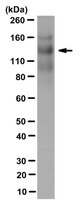
Evaluated by Western Blotting in human thrombospondin-1 purified protein.
Western Blotting Analysis: 0.1 µg/mL of this antibody detected Thrombospondin-1 in 0.1 µg of human thrombospondin-1 purified protein.
Usage Statement
Unless otherwise stated in our catalog or other company documentation accompanying the product(s), our products are intended for research use only and are not to be used for any other purpose, which includes but is not limited to, unauthorized commercial uses, in vitro diagnostic uses, ex vivo or in vivo therapeutic uses or any type of consumption or application to humans or animals.
Storage and Shipping Information
Storage Conditions
Stable for 1 year at -20°C from date of receipt. Handling Recommendations: Upon receipt and prior to removing the cap, centrifuge the vial and gently mix the solution. Aliquot into microcentrifuge tubes and store at -20°C. Avoid repeated freeze/thaw cycles, which may damage IgG and affect product performance.