Wenn Sie das Fenster schließen, wird Ihre Konfiguration nicht gespeichert, es sei denn, Sie haben Ihren Artikel in die Bestellung aufgenommen oder zu Ihren Favoriten hinzugefügt.
Klicken Sie auf OK, um das MILLIPLEX® MAP-Tool zu schließen oder auf Abbrechen, um zu Ihrer Auswahl zurückzukehren.
Wählen Sie konfigurierbare Panels & Premixed-Kits - ODER - Kits für die zelluläre Signaltransduktion & MAPmates™
Konfigurieren Sie Ihre MILLIPLEX® MAP-Kits und lassen sich den Preis anzeigen.
Konfigurierbare Panels & Premixed-Kits
Unser breites Angebot enthält Multiplex-Panels, für die Sie die Analyten auswählen können, die am besten für Ihre Anwendung geeignet sind. Unter einem separaten Register können Sie das Premixed-Cytokin-Format oder ein Singleplex-Kit wählen.
Kits für die zelluläre Signaltransduktion & MAPmates™
Wählen Sie gebrauchsfertige Kits zur Erforschung gesamter Signalwege oder Prozesse. Oder konfigurieren Sie Ihre eigenen Kits mit Singleplex MAPmates™.
Die folgenden MAPmates™ sollten nicht zusammen analysiert werden: -MAPmates™, die einen unterschiedlichen Assaypuffer erfordern. -Phosphospezifische und MAPmate™ Gesamtkombinationen wie Gesamt-GSK3β und Gesamt-GSK3β (Ser 9). -PanTyr und locusspezifische MAPmates™, z.B. Phospho-EGF-Rezeptor und Phospho-STAT1 (Tyr701). -Mehr als 1 Phospho-MAPmate™ für ein einziges Target (Akt, STAT3). -GAPDH und β-Tubulin können nicht mit Kits oder MAPmates™, die panTyr enthalten, analysiert werden.
.
Bestellnummer
Bestellinformationen
St./Pkg.
Liste
Dieser Artikel wurde zu Ihren Favoriten hinzugefügt.
Wählen Sie bitte Spezies, Panelart, Kit oder Probenart
Um Ihr MILLIPLEX® MAP-Kit zu konfigurieren, wählen Sie zunächst eine Spezies, eine Panelart und/oder ein Kit.
Custom Premix Selecting "Custom Premix" option means that all of the beads you have chosen will be premixed in manufacturing before the kit is sent to you.
Catalogue Number
Ordering Description
Qty/Pack
List
Dieser Artikel wurde zu Ihren Favoriten hinzugefügt.
Spezies
Panelart
Gewähltes Kit
Menge
Bestellnummer
Bestellinformationen
St./Pkg.
Listenpreis
96-Well Plate
Menge
Bestellnummer
Bestellinformationen
St./Pkg.
Listenpreis
Weitere Reagenzien hinzufügen (MAPmates erfordern die Verwendung eines Puffer- und Detektionskits)
Menge
Bestellnummer
Bestellinformationen
St./Pkg.
Listenpreis
48-602MAG
Buffer Detection Kit for Magnetic Beads
1 Kit
Platzsparende Option Kunden, die mehrere Kits kaufen, können ihre Multiplex-Assaykomponenten in Kunststoffbeuteln anstelle von Packungen erhalten, um eine kompaktere Lagerung zu ermöglichen.
Dieser Artikel wurde zu Ihren Favoriten hinzugefügt.
Das Produkt wurde in Ihre Bestellung aufgenommen
Sie können nun ein weiteres Kit konfigurieren, ein Premixed-Kit wählen, zur Kasse gehen oder das Bestell-Tool schließen.
Anti-Rabies Virus, clone 509-6, Cat. No. MABF2073, is a mouse monoclonal antibody that detects glycoprotein of Rabies virus, strain CVS-11 and has been tested for use in ELISA, Flow Cytometry, Fluorescence Activated Cell Sorting (FACS), and Neutralizing.
More>>Anti-Rabies Virus, clone 509-6, Cat. No. MABF2073, is a mouse monoclonal antibody that detects glycoprotein of Rabies virus, strain CVS-11 and has been tested for use in ELISA, Flow Cytometry, Fluorescence Activated Cell Sorting (FACS), and Neutralizing. Less<<
SDB (Sicherheitsdatenblätter), Analysenzertifikate und Qualitätszertifikate, Dossiers, Broschüren und andere verfügbare Dokumente.
Glycoprotein (UniProt: O92284) is encoded by the G gene in Rabies virus, strain CVS-11. Glycoprotein is a single-pass type I membrane protein that belongs to the lyssavirus glycoprotein family. It attached the virus to host cellular receptor, inducing endocytosis of the virion. In the endosome, the acidic pH induces conformational changes in the glycoprotein trimer, which trigger fusion between virus and cell membrane. The muscular form of the nicotinic acetylcholine receptor (nAChR), the neuronal cell adhesion molecule (NCAM), and the p75 neurotrophin receptor (p75NTR) are reported to bind glycoprotein and thereby facilitate rabies virus entry into cells. Glycoprotein structure includes a virion surface (aa 20-459), helical region (aa 460-480) and an intravirion (aa 481-524). Arginine 352 is reported to be involved in Rabies virus pathogenicity and its mutation attenuates the virus. Glycoprotein is glycosylated and palmitoylated by host and glycosylation is considered to be crucial for glycoprotein export at the cell surface. Clone 509-6 is shown to neutralize all fixed and street rabies virus strains except some virus isolates from bats. The conformational antigenic site I for this clone is located at aa 231. (Ref.: Wiktor, TJ and Koprowski, H (1980). J. Exp. Med. 152: 99-112; Marissen, WE et al (2005). J. Virol. 79(8): 4672 4678).
References
Product Information
Format
Purified
HS Code
3002 15 90
Presentation
Purified mouse monoclonal antibody IgG2a in PBS without azide.
Anti-Rabies Virus, clone 509-6, Cat. No. MABF2073, is a mouse monoclonal antibody that detects glycoprotein of Rabies virus, strain CVS-11 and has been tested for use in ELISA, Flow Cytometry, Fluorescence Activated Cell Sorting (FACS), and Neutralizing.
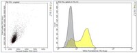
Clone 509-6 detected glycoprotein from Rabies virus, strain CVS-11 in L929 cells expressing the rabies protein. It did not detect glycoprotein in wild type L929 cells.
Evaluated by Flow Cytometry in L929 cells expressing the rabies protein and in L929 (WT) cells.
Flow Cytometry Analysis: 1 µg of this antibody detected glycoprotein of Rabies virus in one million L929 cells expressing the rabies protein.
Usage Statement
Unless otherwise stated in our catalog or other company documentation accompanying the product(s), our products are intended for research use only and are not to be used for any other purpose, which includes but is not limited to, unauthorized commercial uses, in vitro diagnostic uses, ex vivo or in vivo therapeutic uses or any type of consumption or application to humans or animals.
Storage and Shipping Information
Storage Conditions
Stable for 1 year at -20°C from date of receipt. Handling Recommendations: Upon receipt and prior to removing the cap, centrifuge the vial and gently mix the solution. Aliquot into microcentrifuge tubes and store at -20°C. Avoid repeated freeze/thaw cycles, which may damage IgG and affect product performance.