Wenn Sie das Fenster schließen, wird Ihre Konfiguration nicht gespeichert, es sei denn, Sie haben Ihren Artikel in die Bestellung aufgenommen oder zu Ihren Favoriten hinzugefügt.
Klicken Sie auf OK, um das MILLIPLEX® MAP-Tool zu schließen oder auf Abbrechen, um zu Ihrer Auswahl zurückzukehren.
Wählen Sie konfigurierbare Panels & Premixed-Kits - ODER - Kits für die zelluläre Signaltransduktion & MAPmates™
Konfigurieren Sie Ihre MILLIPLEX® MAP-Kits und lassen sich den Preis anzeigen.
Konfigurierbare Panels & Premixed-Kits
Unser breites Angebot enthält Multiplex-Panels, für die Sie die Analyten auswählen können, die am besten für Ihre Anwendung geeignet sind. Unter einem separaten Register können Sie das Premixed-Cytokin-Format oder ein Singleplex-Kit wählen.
Kits für die zelluläre Signaltransduktion & MAPmates™
Wählen Sie gebrauchsfertige Kits zur Erforschung gesamter Signalwege oder Prozesse. Oder konfigurieren Sie Ihre eigenen Kits mit Singleplex MAPmates™.
Die folgenden MAPmates™ sollten nicht zusammen analysiert werden: -MAPmates™, die einen unterschiedlichen Assaypuffer erfordern. -Phosphospezifische und MAPmate™ Gesamtkombinationen wie Gesamt-GSK3β und Gesamt-GSK3β (Ser 9). -PanTyr und locusspezifische MAPmates™, z.B. Phospho-EGF-Rezeptor und Phospho-STAT1 (Tyr701). -Mehr als 1 Phospho-MAPmate™ für ein einziges Target (Akt, STAT3). -GAPDH und β-Tubulin können nicht mit Kits oder MAPmates™, die panTyr enthalten, analysiert werden.
.
Bestellnummer
Bestellinformationen
St./Pkg.
Liste
Dieser Artikel wurde zu Ihren Favoriten hinzugefügt.
Wählen Sie bitte Spezies, Panelart, Kit oder Probenart
Um Ihr MILLIPLEX® MAP-Kit zu konfigurieren, wählen Sie zunächst eine Spezies, eine Panelart und/oder ein Kit.
Custom Premix Selecting "Custom Premix" option means that all of the beads you have chosen will be premixed in manufacturing before the kit is sent to you.
Catalogue Number
Ordering Description
Qty/Pack
List
Dieser Artikel wurde zu Ihren Favoriten hinzugefügt.
Spezies
Panelart
Gewähltes Kit
Menge
Bestellnummer
Bestellinformationen
St./Pkg.
Listenpreis
96-Well Plate
Menge
Bestellnummer
Bestellinformationen
St./Pkg.
Listenpreis
Weitere Reagenzien hinzufügen (MAPmates erfordern die Verwendung eines Puffer- und Detektionskits)
Menge
Bestellnummer
Bestellinformationen
St./Pkg.
Listenpreis
48-602MAG
Buffer Detection Kit for Magnetic Beads
1 Kit
Platzsparende Option Kunden, die mehrere Kits kaufen, können ihre Multiplex-Assaykomponenten in Kunststoffbeuteln anstelle von Packungen erhalten, um eine kompaktere Lagerung zu ermöglichen.
Dieser Artikel wurde zu Ihren Favoriten hinzugefügt.
Das Produkt wurde in Ihre Bestellung aufgenommen
Sie können nun ein weiteres Kit konfigurieren, ein Premixed-Kit wählen, zur Kasse gehen oder das Bestell-Tool schließen.
ABE440
Sigma-AldrichAnti-RAG-1 Antibody
Detect RAG-1 using this rabbit polyclonal antibody, Anti-RAG-1 Antibody validated for use in western blotting, ICC & IP.
More>>Detect RAG-1 using this rabbit polyclonal antibody, Anti-RAG-1 Antibody validated for use in western blotting, ICC & IP. Less<<
Anti-RAG-1 Antibody: SDB (Sicherheitsdatenblätter), Analysenzertifikate und Qualitätszertifikate, Dossiers, Broschüren und andere verfügbare Dokumente.
V(D)J recombination-activating protein 1 (RAG-1) is also called RING finger protein 74. RAG-1 is a component of the RAG complex, which mediates the DNA cleavage phase during V(D)J recombination. As part of the RAG complex, RAG1 mediates the DNA-binding and catalyzes the DNA cleavage activities by introducing a double-strand breaks between the recombination signal sequences (RSS) and the adjacent coding segment. RAG-1 also acts as a E3 ubiquitin-protein ligase that mediates monoubiquitination of histone H3 as well as polyubiquitination of KPNA1. RAG-1 is expressed in maturing lymphoid cells. RAG-1 defects can cause disease states such as: combined cellular and humoral immune defects with granulomas (CHIDG), severe combined immunodeficiency autosomal recessive T-cell-negative/B-cell-negative/NK-cell-positive (T-B-NK+ SCID), Omenn syndrome (OS), and alpha/beta T-cell lymphopenia with gamma/delta T-cell expansion severe cytomegalovirus infection and autoimmunity (T-CMVA).
References
Product Information
Format
Affinity Purified
Presentation
Purified rabbit polyclonal in buffer containing 0.1 M Tris-Glycine (pH 7.4), 150 mM NaCl with 0.05% sodium azide.
Detect RAG-1 using this rabbit polyclonal antibody, Anti-RAG-1 Antibody validated for use in western blotting, ICC & IP.
Key Applications
Western Blotting
Immunocytochemistry
Immunoprecipitation
Application Notes
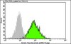
Western Blotting Analysis: 1 µg/mL from a representative lot detected RAG-1 immunopreciptated from 500 µg of HL-60 cell lysate. (Note: It is recommended to immunoprecipitate RAG-1 before performing WB).
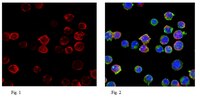
Immunocytochemistry Analysis: 1:100 dilution from a representative lot detected RAG-1 in Jurkat cells.
DyLight® is a registered trademark of Thermo Fisher Scientific.
Biological Information
Immunogen
KLH-conjugated linear peptide corresponding to a region near the N-terminus of human RAG-1.
Concentration
Please refer to the Certificate of Analysis for the lot-specific concentration.
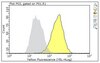
Evaluated by Immunoprecipitation in HL-60 cell lysate.
Immunoprecipitation Analysis: 10 µg of this antibody immunoprecipitated RAG-1 in 500 µg of HL-60 cell lysate. Immunopreciptated protein was then probed with 1 µg/mL of the same antibody in WB.
Usage Statement
Unless otherwise stated in our catalog or other company documentation accompanying the product(s), our products are intended for research use only and are not to be used for any other purpose, which includes but is not limited to, unauthorized commercial uses, in vitro diagnostic uses, ex vivo or in vivo therapeutic uses or any type of consumption or application to humans or animals.