Millipore Sigma Vibrant Logo

ST1202 Anti-PGC-1α Mouse mAb (4C1.3)

Übersicht

Replacement Information

Key Spec Table

Species ReactivityHostAntibody Type
H, M, RMMonoclonal Antibody

Preis & Verfügbarkeit

Bestellnummer VerfügbarkeitVerpackung St./Pkg. Preis Menge
ST1202-1SET
Verfügbarkeit wird abgerufen...
Eingeschränkte Verfügbarkeit
Lieferbar 
Produkt wurde eingestellt
Begrenzter Lagerbestand
Bestätigung der Verfügbarkeit erforderlich
    Restmenge: Angebot folgt
      Restmenge: Angebot folgt
      Bitte erfragen
      Kontakt zum Kundenservice
      Contact Customer Service

      Kst.-Ampulle 1 set
      Preis wird abgerufen...
      Preis nicht abrufbar
      Die Mindestmenge muss ein Vielfaches sein von
      Maximum Quantity is
      Bei Bestätigung Weitere Informationen
      Sie haben () gespart
       
      Bitte erfragen
      Description
      OverviewRecognizes the endogenous forms of PGC-1α, which includes the ~113 kDa PGC-1α protein and the ~38 kDa splice variant, in brown adipose tissue, liver, and kidney.
      Catalogue NumberST1202
      Brand Family Calbiochem®
      SynonymsAnti-Peroxisome Proliferator-Activated Receptor- γ Coactivator 1 α
      Application Data
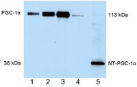
      Detection of human PGC-1α by immunoblottiong. Samples: Whole cell lysates from COS cells transformed with wild-type PGC-1α (5 µg, lane 1; 10 µg, lane 2; 20 µg, lane 3), whole cell extract from brown adipose tissue of a cold-exposed mouse (lane 4), and whole cell lysate from CHO cells transformed with NT-PGC-1α (0.5 µg, lane 5). Primary antibody: Anti-PGC-1α Mouse mAb (4C1.3) (Cat. No. ST1202) (1 µg/ml). Detection chemiluminescence.

      Detection of mouse PGC-1α by immunoblottiong. Samples: NT-PGC-1α Positive Control, (lane 1), whole cell extract from brown adipose tissue of mice kept at room temperature (50 µg; lanes 2-4), whole cell extract from brown adipose tissue of mice exposed to 4°C for 5 h (50 µg; lanes 5-7), and PGC-1α positive control (lane 8). Primary antibody: Anti-PGC-1α Mouse mAb (4C1.3) (Cat. No. ST1202) (1 µg/ml). Detection: chemiluminescence.

      Detection of mouse PGC-1α by immunoblottiong. Samples: PGC-1α positive control (lane 1), nuclear extract from mouse liver (75 µg, lane 2), whole cell extract from mouse kidney (100 µg, lane 3), and whole cell extract from kidney of a PGC-1α knock-out mouse (100 µg, lane 4). Primary antibody: Anti-PGC-1α Mouse mAb (4C1.3) (Cat. No. ST1202) (1 µg/ml). Detection: chemiluminescence.
      References
      ReferencesLai, L., et al. 2008. Genes Dev. 14, 1948.
      Rodgers, J.T., et al. 2008. FEBS Lett. 582, 46.
      Mazzucotelli, A., et al. 2007. Diabetes 10, 2467.
      Nemoto, S., et al. 2005. J. Biol. Chem. 16, 16456.
      Product Information
      FormLiquid
      FormulationOne vial of 100 µg antibody in 50 mM PBS, see vial for lot-specific concentration. One vial of NT-PGC-1α Positive Control, supplied as 25 µl whole cell extract in RIPA buffer containing 10 mM &beta-mercaptoethanol and 1% SDS; load 10 µl per lane; add sample buffer prior to SDS-PAGE loading.
      PreservativeNone
      Quality LevelMQ100
      Applications
      Application ReferencesParaffin Sections
      Koh, Y.J., et al. 2009. Exp. Mol. Med. 41, 880.
      Key Applications Immunoblotting (Western Blotting)
      Immunocytochemistry
      Immunoprecipitation
      Paraffin Sections
      Application NotesImmunoblotting (1 µg/ml)
      Immunocytochemistry (5-10 µg/ml)
      Immunoprecipitation (10 µg/ml)
      Paraffin Sections (whole mount) (see application references)
      Application CommentsEndogenous PGC-1α is found primarily in the nucleus and NT-PGC-1α is found in both cytosolic and nuclear fractions. Antibody should be titrated for optimal results in individual systems.
      Biological Information
      Immunogena recombinant protein consisting of amino acids 1-120 of mouse PGC-1α
      ImmunogenMouse
      Clone4C1.3
      HostMouse
      IsotypeIgG
      Species Reactivity
      • Human
      • Mouse
      • Rat
      Antibody TypeMonoclonal Antibody
      Physicochemical Information
      Dimensions
      Materials Information
      Toxicological Information
      Safety Information according to GHS
      Safety Information
      R PhraseR: 21/22-36/37/38

      Harmful in contact with skin and if swallowed.
      Irritating to eyes, respiratory system and skin.
      S PhraseS: 26-36/37-45

      In case of contact with eyes, rinse immediately with plenty of water and seek medical advice.
      Wear suitable protective clothing and gloves.
      In case of accident or if you feel unwell, seek medical advice immediately (show the label where possible).
      Product Usage Statements
      Storage and Shipping Information
      Ship Code Dry Ice Only
      Toxicity Multiple Toxicity Values, refer to MSDS
      Storage ≤ -70°C
      Avoid freeze/thaw Avoid freeze/thaw
      Do not freeze Ok to freeze
      Special InstructionsFollowing initial thaw, aliquot and freeze (-70°C).
      Packaging Information
      Transport Information
      Supplemental Information
      Specifications
      Global Trade Item Number
      Bestellnummer GTIN
      ST1202-1SET 04055977224085

      Documentation

      Anti-PGC-1α Mouse mAb (4C1.3) SDB

      Titel

      Sicherheitsdatenblatt (SDB) 

      Anti-PGC-1α Mouse mAb (4C1.3) Analysenzertifikate

      TitelChargennummer
      ST1202

      Literatur

      Übersicht
      Lai, L., et al. 2008. Genes Dev. 14, 1948.
      Rodgers, J.T., et al. 2008. FEBS Lett. 582, 46.
      Mazzucotelli, A., et al. 2007. Diabetes 10, 2467.
      Nemoto, S., et al. 2005. J. Biol. Chem. 16, 16456.

      Literaturstellen

      Titel
    • Chang, J.S., et al. 2010. J. Biol. Chem. In Press.
    • Estall, J. L., et al. 2009. Diabetes 58, 1499.
    • Zhang Y., et al. 2009. J. Biol. Chem.) in Press.

    • Paraffin Sections
      Koh, Y.J., et al. 2009. Exp. Mol. Med. 41, 880.
    • Datenblatt

      Note that this data sheet is not lot-specific and is representative of the current specifications for this product. Please consult the vial label and the certificate of analysis for information on specific lots. Also note that shipping conditions may differ from storage conditions.

      Revision23-October-2018 JSW
      SynonymsAnti-Peroxisome Proliferator-Activated Receptor- γ Coactivator 1 α
      ApplicationImmunoblotting (1 µg/ml)
      Immunocytochemistry (5-10 µg/ml)
      Immunoprecipitation (10 µg/ml)
      Paraffin Sections (whole mount) (see application references)
      Application Data
      Detection of human PGC-1α by immunoblottiong. Samples: Whole cell lysates from COS cells transformed with wild-type PGC-1α (5 µg, lane 1; 10 µg, lane 2; 20 µg, lane 3), whole cell extract from brown adipose tissue of a cold-exposed mouse (lane 4), and whole cell lysate from CHO cells transformed with NT-PGC-1α (0.5 µg, lane 5). Primary antibody: Anti-PGC-1α Mouse mAb (4C1.3) (Cat. No. ST1202) (1 µg/ml). Detection chemiluminescence.

      Detection of mouse PGC-1α by immunoblottiong. Samples: NT-PGC-1α Positive Control, (lane 1), whole cell extract from brown adipose tissue of mice kept at room temperature (50 µg; lanes 2-4), whole cell extract from brown adipose tissue of mice exposed to 4°C for 5 h (50 µg; lanes 5-7), and PGC-1α positive control (lane 8). Primary antibody: Anti-PGC-1α Mouse mAb (4C1.3) (Cat. No. ST1202) (1 µg/ml). Detection: chemiluminescence.

      Detection of mouse PGC-1α by immunoblottiong. Samples: PGC-1α positive control (lane 1), nuclear extract from mouse liver (75 µg, lane 2), whole cell extract from mouse kidney (100 µg, lane 3), and whole cell extract from kidney of a PGC-1α knock-out mouse (100 µg, lane 4). Primary antibody: Anti-PGC-1α Mouse mAb (4C1.3) (Cat. No. ST1202) (1 µg/ml). Detection: chemiluminescence.
      DescriptionProtein G purified mouse monclonal antibody. Recognizes the endogenous forms of PGC-1α, which includes the ~113 kDa PGC-1α protein and the ~38 kDa splice variant.
      BackgroundPGC-1α is a co-activator of PPARα, PPARγ, and other transcription factors and regulates the transcriptonal program of adaptive thermogenesis in brown adipose tissue, hepatic/renal gluconeogenesis, and muscle fiber type switching. The full-length protein is 113 kDa and is induced in brown adipose tissue by cold exposure, in liver and kidney by fasting, and in skeletal muscle by exercise. Alternative splicing of the full-length gene produces a 270 aa N-terminal splice variant that migrates at ~38 kDa and is induced by the same physiological signals that induce expression of the full-length protein. The N-terminal PGC-1α splice variant is also subject to post translational modifications that alter its migration and apparent molecular weight.
      HostMouse
      Immunogen speciesMouse
      Immunogena recombinant protein consisting of amino acids 1-120 of mouse PGC-1α
      Clone4C1.3
      IsotypeIgG
      Specieshuman, mouse, rat
      FormLiquid
      FormulationOne vial of 100 µg antibody in 50 mM PBS, see vial for lot-specific concentration. One vial of NT-PGC-1α Positive Control, supplied as 25 µl whole cell extract in RIPA buffer containing 10 mM &beta-mercaptoethanol and 1% SDS; load 10 µl per lane; add sample buffer prior to SDS-PAGE loading.
      PreservativeNone
      CommentsEndogenous PGC-1α is found primarily in the nucleus and NT-PGC-1α is found in both cytosolic and nuclear fractions. Antibody should be titrated for optimal results in individual systems.
      Storage Avoid freeze/thaw
      ≤ -70°C
      Do Not Freeze Ok to freeze
      Special InstructionsFollowing initial thaw, aliquot and freeze (-70°C).
      Toxicity Multiple Toxicity Values, refer to MSDS
      ReferencesLai, L., et al. 2008. Genes Dev. 14, 1948.
      Rodgers, J.T., et al. 2008. FEBS Lett. 582, 46.
      Mazzucotelli, A., et al. 2007. Diabetes 10, 2467.
      Nemoto, S., et al. 2005. J. Biol. Chem. 16, 16456.
      Citation
    • Chang, J.S., et al. 2010. J. Biol. Chem. In Press.
    • Estall, J. L., et al. 2009. Diabetes 58, 1499.
    • Zhang Y., et al. 2009. J. Biol. Chem.) in Press.

    • Paraffin Sections
      Koh, Y.J., et al. 2009. Exp. Mol. Med. 41, 880.
    • Application referencesParaffin Sections
      Koh, Y.J., et al. 2009. Exp. Mol. Med. 41, 880.