Wenn Sie das Fenster schließen, wird Ihre Konfiguration nicht gespeichert, es sei denn, Sie haben Ihren Artikel in die Bestellung aufgenommen oder zu Ihren Favoriten hinzugefügt.
Klicken Sie auf OK, um das MILLIPLEX® MAP-Tool zu schließen oder auf Abbrechen, um zu Ihrer Auswahl zurückzukehren.
Wählen Sie konfigurierbare Panels & Premixed-Kits - ODER - Kits für die zelluläre Signaltransduktion & MAPmates™
Konfigurieren Sie Ihre MILLIPLEX® MAP-Kits und lassen sich den Preis anzeigen.
Konfigurierbare Panels & Premixed-Kits
Unser breites Angebot enthält Multiplex-Panels, für die Sie die Analyten auswählen können, die am besten für Ihre Anwendung geeignet sind. Unter einem separaten Register können Sie das Premixed-Cytokin-Format oder ein Singleplex-Kit wählen.
Kits für die zelluläre Signaltransduktion & MAPmates™
Wählen Sie gebrauchsfertige Kits zur Erforschung gesamter Signalwege oder Prozesse. Oder konfigurieren Sie Ihre eigenen Kits mit Singleplex MAPmates™.
Die folgenden MAPmates™ sollten nicht zusammen analysiert werden: -MAPmates™, die einen unterschiedlichen Assaypuffer erfordern. -Phosphospezifische und MAPmate™ Gesamtkombinationen wie Gesamt-GSK3β und Gesamt-GSK3β (Ser 9). -PanTyr und locusspezifische MAPmates™, z.B. Phospho-EGF-Rezeptor und Phospho-STAT1 (Tyr701). -Mehr als 1 Phospho-MAPmate™ für ein einziges Target (Akt, STAT3). -GAPDH und β-Tubulin können nicht mit Kits oder MAPmates™, die panTyr enthalten, analysiert werden.
.
Bestellnummer
Bestellinformationen
St./Pkg.
Liste
Dieser Artikel wurde zu Ihren Favoriten hinzugefügt.
Wählen Sie bitte Spezies, Panelart, Kit oder Probenart
Um Ihr MILLIPLEX® MAP-Kit zu konfigurieren, wählen Sie zunächst eine Spezies, eine Panelart und/oder ein Kit.
Custom Premix Selecting "Custom Premix" option means that all of the beads you have chosen will be premixed in manufacturing before the kit is sent to you.
Catalogue Number
Ordering Description
Qty/Pack
List
Dieser Artikel wurde zu Ihren Favoriten hinzugefügt.
Spezies
Panelart
Gewähltes Kit
Menge
Bestellnummer
Bestellinformationen
St./Pkg.
Listenpreis
96-Well Plate
Menge
Bestellnummer
Bestellinformationen
St./Pkg.
Listenpreis
Weitere Reagenzien hinzufügen (MAPmates erfordern die Verwendung eines Puffer- und Detektionskits)
Menge
Bestellnummer
Bestellinformationen
St./Pkg.
Listenpreis
48-602MAG
Buffer Detection Kit for Magnetic Beads
1 Kit
Platzsparende Option Kunden, die mehrere Kits kaufen, können ihre Multiplex-Assaykomponenten in Kunststoffbeuteln anstelle von Packungen erhalten, um eine kompaktere Lagerung zu ermöglichen.
Dieser Artikel wurde zu Ihren Favoriten hinzugefügt.
Das Produkt wurde in Ihre Bestellung aufgenommen
Sie können nun ein weiteres Kit konfigurieren, ein Premixed-Kit wählen, zur Kasse gehen oder das Bestell-Tool schließen.
Anti-PD-L1, clone 5H1, Cat. No. MABC1115, is a mouse monoclonal antibody that detects PD-L1 and has been tested for use in Immunocytochemistry, Immunohistochemistry (Paraffin), and Western Blotting.
More>>Anti-PD-L1, clone 5H1, Cat. No. MABC1115, is a mouse monoclonal antibody that detects PD-L1 and has been tested for use in Immunocytochemistry, Immunohistochemistry (Paraffin), and Western Blotting. Less<<
Programmed cell death 1 ligand 1 (UniProt Q9NZQ7; also known as B7-H1, B7 homolog 1, CD274, PD-L1, PDCD1 ligand 1, Programmed death ligand 1) is encoded by the CD274 (also known as B7H1, PDCD1L1, PDCD1LG1, PDL1) gene (Gene ID 29126) in human. PD-L1 is a single-pass type I membrane protein that plays a critical role in induction and maintenance of immune tolerance to self. It is expressed on activated T- and B-cells, dendritic cells, keratinocytes, and monocytes. Higher expression has also been reported in heart, skeletal muscle, placenta, and lung. PD-1 and PD-1 ligands 1 and 2 (PD-L1 and PD-L2) are B7:CD28 family members that regulate T cell activation and peripheral tolerance. When engaged together with the TCR, the interaction of PD-1 with its ligands delivers an inhibitory signal to T cell proliferation and cytokine production. While PD-L1 is broadly expressed in hematopoietic and nonhematopoietic cells, PD-L2 expression is highly restricted to antigen presenting cells (APCs), including dendritic cells (DCs) and macrophages. The PD-1 pathway plays a key role in the progressive loss of effector T cell responses during chronic HIV infection. Under some conditions, blockade of this pathway can restore many T cell functions. PD-L1 is initially produced with signal peptide (aa 1-18) sequence, which is subsequently cleaved off to produce the mature protein with a large extracellular (aa 19-238) region that contains an Ig-like V-type domain (aa 19-127) and an Ig-like C2-type domain (aa 133-225), followed by a transmembrane domain (aa 239-259) and a cytoplasmic tail (aa 260-290).
References
Product Information
Format
Purified
Presentation
Purified mouse monoclonal antibody IgG1 in PBS without azide.
Applications
Application
Anti-PD-L1, clone 5H1, Cat. No. MABC1115, is a mouse monoclonal antibody that detects PD-L1 and has been tested for use in Immunocytochemistry, Immunohistochemistry (Paraffin), and Western Blotting.
Key Applications
Immunocytochemistry
Immunohistochemistry (Paraffin)
Western Blotting
Application Notes
Immunohistochemistry (Paraffin) Analysis: A representative lot detected PD-L1 in Immunohistochemistry applications (Bigelow, E., et. al. (2013). J Vis Exp. 3;(71); Spranger, S., et. al. (2013). Sci Transl Med. 5(200):200ra116; Parra, E.R., et. al. (2018). Appl Immunohistochem Mol Morphol. 26(2):83-93; Andorsky, D.J., et. al. (2011). Clin Cancer Res. 17(13):4232-44; Sunshine, J.C., et. al. (2017). Clin Cancer Res. 23(16):4938-4944).
Western Blotting Analysis: A representative lot detected PD-L1 in Western Blotting applications (Parra, E.R., et. al. (2018). Appl Immunohistochem. Mol. Morphol. 26(2):83-93_.
Biological Information
Immunogen
Full-length human recombinant PD-L1.
Epitope
extracellular domain
Clone
5H1
Concentration
Please refer to lot specific datasheet.
Host
Mouse
Specificity
Clone 5H1 detects human Programmed cell death ligand 1 (PD-L1). It targets an epitope with in the extracellular domain.
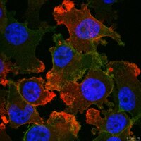
Evaluated by Immunocytochemistry in MDA-MB-231 cells.
Immunocytochemistry Analysis: A 1:50 dilution of this antibody detected PD-L1 in MDA-MB-231 cells.
Usage Statement
Unless otherwise stated in our catalog or other company documentation accompanying the product(s), our products are intended for research use only and are not to be used for any other purpose, which includes but is not limited to, unauthorized commercial uses, in vitro diagnostic uses, ex vivo or in vivo therapeutic uses or any type of consumption or application to humans or animals.
Storage and Shipping Information
Storage Conditions
Stable for 1 year at -20°C from date of receipt. Handling Recommendations: Upon receipt and prior to removing the cap, centrifuge the vial and gently mix the solution. Aliquot into microcentrifuge tubes and store at -20°C. Avoid repeated freeze/thaw cycles, which may damage IgG and affect product performance.