Wenn Sie das Fenster schließen, wird Ihre Konfiguration nicht gespeichert, es sei denn, Sie haben Ihren Artikel in die Bestellung aufgenommen oder zu Ihren Favoriten hinzugefügt.
Klicken Sie auf OK, um das MILLIPLEX® MAP-Tool zu schließen oder auf Abbrechen, um zu Ihrer Auswahl zurückzukehren.
Wählen Sie konfigurierbare Panels & Premixed-Kits - ODER - Kits für die zelluläre Signaltransduktion & MAPmates™
Konfigurieren Sie Ihre MILLIPLEX® MAP-Kits und lassen sich den Preis anzeigen.
Konfigurierbare Panels & Premixed-Kits
Unser breites Angebot enthält Multiplex-Panels, für die Sie die Analyten auswählen können, die am besten für Ihre Anwendung geeignet sind. Unter einem separaten Register können Sie das Premixed-Cytokin-Format oder ein Singleplex-Kit wählen.
Kits für die zelluläre Signaltransduktion & MAPmates™
Wählen Sie gebrauchsfertige Kits zur Erforschung gesamter Signalwege oder Prozesse. Oder konfigurieren Sie Ihre eigenen Kits mit Singleplex MAPmates™.
Die folgenden MAPmates™ sollten nicht zusammen analysiert werden: -MAPmates™, die einen unterschiedlichen Assaypuffer erfordern. -Phosphospezifische und MAPmate™ Gesamtkombinationen wie Gesamt-GSK3β und Gesamt-GSK3β (Ser 9). -PanTyr und locusspezifische MAPmates™, z.B. Phospho-EGF-Rezeptor und Phospho-STAT1 (Tyr701). -Mehr als 1 Phospho-MAPmate™ für ein einziges Target (Akt, STAT3). -GAPDH und β-Tubulin können nicht mit Kits oder MAPmates™, die panTyr enthalten, analysiert werden.
.
Bestellnummer
Bestellinformationen
St./Pkg.
Liste
Dieser Artikel wurde zu Ihren Favoriten hinzugefügt.
Wählen Sie bitte Spezies, Panelart, Kit oder Probenart
Um Ihr MILLIPLEX® MAP-Kit zu konfigurieren, wählen Sie zunächst eine Spezies, eine Panelart und/oder ein Kit.
Custom Premix Selecting "Custom Premix" option means that all of the beads you have chosen will be premixed in manufacturing before the kit is sent to you.
Catalogue Number
Ordering Description
Qty/Pack
List
Dieser Artikel wurde zu Ihren Favoriten hinzugefügt.
Spezies
Panelart
Gewähltes Kit
Menge
Bestellnummer
Bestellinformationen
St./Pkg.
Listenpreis
96-Well Plate
Menge
Bestellnummer
Bestellinformationen
St./Pkg.
Listenpreis
Weitere Reagenzien hinzufügen (MAPmates erfordern die Verwendung eines Puffer- und Detektionskits)
Menge
Bestellnummer
Bestellinformationen
St./Pkg.
Listenpreis
48-602MAG
Buffer Detection Kit for Magnetic Beads
1 Kit
Platzsparende Option Kunden, die mehrere Kits kaufen, können ihre Multiplex-Assaykomponenten in Kunststoffbeuteln anstelle von Packungen erhalten, um eine kompaktere Lagerung zu ermöglichen.
Dieser Artikel wurde zu Ihren Favoriten hinzugefügt.
Das Produkt wurde in Ihre Bestellung aufgenommen
Sie können nun ein weiteres Kit konfigurieren, ein Premixed-Kit wählen, zur Kasse gehen oder das Bestell-Tool schließen.
Anti-Integrin aV, clone LM142, Cat. No. MAB1978-I, is a mouse monoclonal antibody that detects Integrin aV and is used in Affinity Binding Assay, Flow Cytometry, Immunocytochemistry, Immunofluorescence, Immunoprecipitation, and Surface plasmon resonance.
More>>Anti-Integrin aV, clone LM142, Cat. No. MAB1978-I, is a mouse monoclonal antibody that detects Integrin aV and is used in Affinity Binding Assay, Flow Cytometry, Immunocytochemistry, Immunofluorescence, Immunoprecipitation, and Surface plasmon resonance. Less<<
Integrin aV (UniProt: P06756; also known as Vitronectin receptor, Vitronectin receptor subunit alpha, CD51) is encoded by the ITGAV (also known as MSK8, VNRA, VNTR) gene (Gene ID: 3685) in human. Integrins are heterodimeric integral membrane proteins composed of an a subunit and a b subunit that function in cell surface adhesion and signaling. They contain a large extracellular domain responsible for ligand binding, a single transmembrane domain, and a cytoplasmic domain. The exact combination of various a- and b-subunits dictates the binding specificity of integrins to different ECM components. Although both subunits are required for adhesion, the binding specificity primarily depends on the extracellular portion of the a-subunit. The structural and functional diversity of the integrin family is based upon the pairing abilities of the individual a and b subunits. Integrins are not constitutively active and their activation from a low ligand-binding affinity state to high ligand-binding affinity state requires conformational change that can originate from either from their cytoplasmic or extracellular interactions. The recognition site for most integrins that bind the ECM consists of an RGD (arginine-glycine-aspartic acid) sequence. Integrin aV is a single-pass membrane glycoprotein that is synthesized with a signal peptide (aa 1-30), which is subsequently cleaved off to generate the mature form. The mature form contains an extracellular domain (aa 31-992), a transmembrane domain (aa 993-1016), and a cytoplasmic domain (aa 1017-1048). The a-subunit is composed of a heavy (aa 31-889) and a light (aa 891-1048) chain linked by a disulfide bond. Integrin aV can associate with b-1, b-3, b -5, b-6, or b-8 in a non-covalent manner. (Ref.: Cheresh, DA. (1987). Proc. Natl. Acad. Sci. USA. 84(18); 6471-6475).
References
Product Information
Format
Purified
Presentation
Purified mouse monoclonal antibody IgG1 in buffer containing 0.1 M Tris-Glycine (pH 7.4), 150 mM NaCl with 0.05% sodium azide.
Anti-Integrin aV, clone LM142, Cat. No. MAB1978-I, is a mouse monoclonal antibody that detects Integrin aV and is used in Affinity Binding Assay, Flow Cytometry, Immunocytochemistry, Immunofluorescence, Immunoprecipitation, and Surface plasmon resonance.
Key Applications
Affinity Binding Assay
Flow Cytometry
Immunocytochemistry
Immunofluorescence
Immunoprecipitation
Surface plasmon resonance
Application Notes
Tested Applications
Surface plasmon resonance: A representative lot detected Integrin aV in Surface plasmon resonance applications (Chesnokova, L.S., et. al. (2011). J Virol. 85(24):13214-23).
Immunoprecipitation Analysis: A representative lot immunoprecipitated Integrin aV in Immunoprecipitation applications (Ueda, M., et. al. (2010). Biomaterials. 31(25):6394-9; Cheresh, D.A., et. al. (1987). Proc Natl Acad Sci USA. 84(18):6471-5).
Immunocytochemistry Analysis: A 1:25 dilution from a representative lot detected Integrin aV in A431 cells.
Immunofluorescence Analysis: A representative lot detected Integrin aV in Immunofluorescence applications (Straub, S., et. al. (2013). PLoS One. 8(1):e53309).
Affinity Binding Assay: A representative lot detected Integrin aV in Affinity Binding Assay applications (Kapp, T.G., et. al. (2017). Sci Rep. 7:39805).
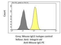
Flow Cytometry Analysis: A representative lot detected Integrin aV in Flow Cytometry applications (Borbely, A., et. al. (2019). Pharmaceutics. 11(4):151).
Immunocytochemistry Analysis: A representative lot detected Integrin aV in Immunocytochemistry applications (Cheresh, D.A., et. al. (1987). Proc Natl Acad Sci USA. 84(18):6471-5).
Note: Actual optimal working dilutions must be determined by end user as specimens, and experimental conditions may vary with the end user
Biological Information
Immunogen
Purified aVb3 isolated from human placenta.
Epitope
Unknown
Clone
LM142
Concentration
0.5 mg/mL. Please refer to guidance on suggested starting dilutions and/or titers per application and sample type.
Host
Mouse
Specificity
Clone LM142 is a mouse monoclonal antibody that detects human Integrin aV.
Flow Cytometry Analysis: 1 µg of this antibody detected Integrin aV in one million HeLa cells.
Usage Statement
Unless otherwise stated in our catalog or other company documentation accompanying the product(s), our products are intended for research use only and are not to be used for any other purpose, which includes but is not limited to, unauthorized commercial uses, in vitro diagnostic uses, ex vivo or in vivo therapeutic uses or any type of consumption or application to humans or animals.
Storage and Shipping Information
Storage Conditions
Recommend storage at +2°C to +8°C. For long term storage antibodies can be kept at -20°C. Avoid repeated freeze-thaws.