Wenn Sie das Fenster schließen, wird Ihre Konfiguration nicht gespeichert, es sei denn, Sie haben Ihren Artikel in die Bestellung aufgenommen oder zu Ihren Favoriten hinzugefügt.
Klicken Sie auf OK, um das MILLIPLEX® MAP-Tool zu schließen oder auf Abbrechen, um zu Ihrer Auswahl zurückzukehren.
Wählen Sie konfigurierbare Panels & Premixed-Kits - ODER - Kits für die zelluläre Signaltransduktion & MAPmates™
Konfigurieren Sie Ihre MILLIPLEX® MAP-Kits und lassen sich den Preis anzeigen.
Konfigurierbare Panels & Premixed-Kits
Unser breites Angebot enthält Multiplex-Panels, für die Sie die Analyten auswählen können, die am besten für Ihre Anwendung geeignet sind. Unter einem separaten Register können Sie das Premixed-Cytokin-Format oder ein Singleplex-Kit wählen.
Kits für die zelluläre Signaltransduktion & MAPmates™
Wählen Sie gebrauchsfertige Kits zur Erforschung gesamter Signalwege oder Prozesse. Oder konfigurieren Sie Ihre eigenen Kits mit Singleplex MAPmates™.
Die folgenden MAPmates™ sollten nicht zusammen analysiert werden: -MAPmates™, die einen unterschiedlichen Assaypuffer erfordern. -Phosphospezifische und MAPmate™ Gesamtkombinationen wie Gesamt-GSK3β und Gesamt-GSK3β (Ser 9). -PanTyr und locusspezifische MAPmates™, z.B. Phospho-EGF-Rezeptor und Phospho-STAT1 (Tyr701). -Mehr als 1 Phospho-MAPmate™ für ein einziges Target (Akt, STAT3). -GAPDH und β-Tubulin können nicht mit Kits oder MAPmates™, die panTyr enthalten, analysiert werden.
.
Bestellnummer
Bestellinformationen
St./Pkg.
Liste
Dieser Artikel wurde zu Ihren Favoriten hinzugefügt.
Wählen Sie bitte Spezies, Panelart, Kit oder Probenart
Um Ihr MILLIPLEX® MAP-Kit zu konfigurieren, wählen Sie zunächst eine Spezies, eine Panelart und/oder ein Kit.
Custom Premix Selecting "Custom Premix" option means that all of the beads you have chosen will be premixed in manufacturing before the kit is sent to you.
Catalogue Number
Ordering Description
Qty/Pack
List
Dieser Artikel wurde zu Ihren Favoriten hinzugefügt.
Spezies
Panelart
Gewähltes Kit
Menge
Bestellnummer
Bestellinformationen
St./Pkg.
Listenpreis
96-Well Plate
Menge
Bestellnummer
Bestellinformationen
St./Pkg.
Listenpreis
Weitere Reagenzien hinzufügen (MAPmates erfordern die Verwendung eines Puffer- und Detektionskits)
Menge
Bestellnummer
Bestellinformationen
St./Pkg.
Listenpreis
48-602MAG
Buffer Detection Kit for Magnetic Beads
1 Kit
Platzsparende Option Kunden, die mehrere Kits kaufen, können ihre Multiplex-Assaykomponenten in Kunststoffbeuteln anstelle von Packungen erhalten, um eine kompaktere Lagerung zu ermöglichen.
Dieser Artikel wurde zu Ihren Favoriten hinzugefügt.
Das Produkt wurde in Ihre Bestellung aufgenommen
Sie können nun ein weiteres Kit konfigurieren, ein Premixed-Kit wählen, zur Kasse gehen oder das Bestell-Tool schließen.
09-893
Sigma-AldrichAnti-BAF (BANF1) Antibody
Use Anti-BAF (BANF1) Antibody (Rabbit Polyclonal Antibody) validated in WB to detect BAF (BANF1) also known as 53 kDa BRG1-associated factor A.
More>>Use Anti-BAF (BANF1) Antibody (Rabbit Polyclonal Antibody) validated in WB to detect BAF (BANF1) also known as 53 kDa BRG1-associated factor A. Less<<
Anti-BAF (BANF1) Antibody: SDB (Sicherheitsdatenblätter), Analysenzertifikate und Qualitätszertifikate, Dossiers, Broschüren und andere verfügbare Dokumente.
Barrier-to-auto integration factor (BAF or BANF1) plays fundamental roles in nuclear assembly, chromatin organization, gene expression and development. BAF may potently compress chromatin structure and be involved in membrane recruitment and chromatin decondensation during nuclear assembly. BAF contains 2 non-specific dsDNA-binding sites which may promote DNA cross-bridging. BAF is exploited by retroviruses for inhibiting self-destructing autointegration of retroviral DNA, thereby promoting integration of viral DNA into the host chromosome. BAF is found in both the nucleus and cytoplasm and is significantly enriched at the nuclear inner membrane, diffused throughout the nucleus during interphase and concentrated at the chromosomes during the M-phase.
Use Anti-BAF (BANF1) Antibody (Rabbit Polyclonal Antibody) validated in WB to detect BAF (BANF1) also known as 53 kDa BRG1-associated factor A.
Key Applications
Western Blotting
Application Notes
Western Blot Analysis: A 1:1,000 dilution from a previous lot detected BAF in C2C12, Hek293, HeLa, HepG2, Huvec, Jurkat, L6, NIH/3T3, PC12, PC3, and RAW264.7 cell lysates.
Biological Information
Immunogen
KLH-conjugated linear peptide corresponding to human BAF (BANF1).
The protein encoded by this gene was first identified by its ability to protect retroviruses from intramolecular integration and therefore promote intermolecular integration into the host cell genome. The protein forms a homodimer which localizes to both the nucleus and cytoplasm and is specifically associated with chromosomes during mitosis. This protein binds to double stranded DNA in a non-specific manner and also binds to LEM-domain containing proteins of the nuclear envelope. This protein is thought to facilitate nuclear reassembly by binding with both DNA and inner nuclear membrane proteins and thereby recruit chromatin to the nuclear periphery. Alternative splicing results in multiple transcript variants encoding the same protein.
FUNCTION: Involved in transcriptional activation and repression of select genes by chromatin remodeling (alteration of DNA-nucleosome topology). Required for maximal ATPase activity of SMARCA4/BRG1 and for association of the SMARCA4/BRG1 containing remodeling complex BAF with chromatin/nuclear matrix. Component of the NuA4 histone acetyltransferase (HAT) complex which is involved in transcriptional activation of select genes principally by acetylation of nucleosomal histones H4 and H2A. This modification may both alter nucleosome - DNA interactions and promote interaction of the modified histones with other proteins which positively regulate transcription. This complex may be required for the activation of transcriptional programs associated with oncogene and proto-oncogene mediated growth induction, tumor suppressor mediated growth arrest and replicative senescence, apoptosis, and DNA repair. NuA4 may also play a direct role in DNA repair when recruited to sites of DNA damage. Also involved in vitamin D-coupled transcription regulation via its association with the WINAC complex, a chromatin-remodeling complex recruited by vitamin D receptor (VDR), which is required for the ligand-bound VDR-mediated transrepression of the CYP27B1 gene By similarity. SUBUNIT STRUCTURE: Component of numerous complexes with chromatin remodeling and histone acetyltransferase activity. Component of the NuA4 histone acetyltransferase complex which contains the catalytic subunit KAT5/TIP60 and the subunits EP400, TRRAP/PAF400, BRD8/SMAP, EPC1, DMAP1/DNMAP1, RUVBL1/TIP49, RUVBL2, ING3, actin, ACTL6A/BAF53A, MORF4L1/MRG15, MORF4L2/MRGX, MRGBP, YEATS4/GAS41, VPS72/YL1 and EAF6. The NuA4 complex interacts with MYC and the adenovirus E1A protein. Component of a NuA4-related complex which contains EP400, TRRAP/PAF400, SRCAP, BRD8/SMAP, EPC1, DMAP1/DNMAP1, RUVBL1/TIP49, RUVBL2, actin, ACTL6A/BAF53A, VPS72 and YEATS4/GAS41. Component of the BAF complex, which includes at least actin (ACTB), ARID1A, ARID1B/BAF250, SMARCA2, SMARCA4/BRG1, ACTL6A/BAF53, ACTL6B/BAF53B, SMARCE1/BAF57, SMARCC1/BAF155, SMARCC2/BAF170, SMARCB1/SNF5/INI1, and one or more of SMARCD1/BAF60A, SMARCD2/BAF60B, or SMARCD3/BAF60C. In muscle cells, the BAF complex also contains DPF3. Component of the BAF53 complex, at least composed of ACTL6A/BAF53A, RUVBL1/TIP49, SMARCA2/BRM, and TRRAP/PAF400, and which may also include a HAT activity related to, but distinct from, that of KAT5. ACTL6A interacts with SMARCA4/BRG1. Component of the chromatin-remodeling INO80 complex, at least composed of ACTL6A, ACTR5, ACTR8, RVBL1, RVBL2, INO80, INO80B, INO80C, INO80D and INO80E. Component of the WINAC complex, at least composed of SMARCA2, SMARCA4, SMARCB1, SMARCC1, SMARCC2, SMARCD1, SMARCE1, ACTL6A, BAZ1B/WSTF, ARID1A, SUPT16H, CHAF1A and TOP2B. SUBCELLULAR LOCATION: Nucleus. SEQUENCE SIMILARITIES: Belongs to the actin family.
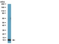
Molecular Weight
~10 kDa
Physicochemical Information
Dimensions
Materials Information
Toxicological Information
Safety Information according to GHS
Safety Information
Product Usage Statements
Quality Assurance
Evaluated by Western Blot in HeLa cell lysate. Western Blot Analysis: A 1:1,000 dilution of this antibody detected BAF in 10 µg of HeLa cell lysate.
Usage Statement
Unless otherwise stated in our catalog or other company documentation accompanying the product(s), our products are intended for research use only and are not to be used for any other purpose, which includes but is not limited to, unauthorized commercial uses, in vitro diagnostic uses, ex vivo or in vivo therapeutic uses or any type of consumption or application to humans or animals.
Storage and Shipping Information
Storage Conditions
Stable for 1 year at -20°C from date of receipt. Handling Recommendations: Upon first thaw, and prior to removing the cap, centrifuge the vial and gently mix the solution. Aliquot into microcentrifuge tubes and store at -20°C. Avoid repeated freeze/thaw cycles, which may damage IgG and affect product performance.
Systems analysis of the prostate tumor suppressor NKX3.1 supports roles in DNA repair and luminal cell differentiation. Yang, CC; Chung, A; Ku, CY; Brill, LM; Williams, R; Wolf, DA F1000Research
3
115
2014
NKX3.1 is a homeobox transcription factor whose function as a prostate tumor suppressor remains insufficiently understood because neither the transcriptional program governed by NKX3.1, nor its interacting proteins have been fully revealed. Using affinity purification and mass spectrometry, we have established an extensive NKX3.1 interactome which contains the DNA repair proteins Ku70, Ku80, and PARP, thus providing a molecular underpinning to previous reports implicating NKX3.1 in DNA repair. Transcriptomic profiling of NKX3.1-negative prostate epithelial cells acutely expressing NKX3.1 revealed a rapid and complex response that is a near mirror image of the gene expression signature of human prostatic intraepithelial neoplasia (PIN). Pathway and network analyses suggested that NKX3.1 actuates a cellular reprogramming toward luminal cell differentiation characterized by suppression of pro-oncogenic c-MYC and interferon-STAT signaling and activation of tumor suppressor pathways. Consistently, ectopic expression of NKX3.1 conferred a growth arrest depending on TNFα and JNK signaling. We propose that the tumor suppressor function of NKX3.1 entails a transcriptional program that maintains the differentiation state of secretory luminal cells and that disruption of NKX3.1 contributes to prostate tumorigenesis by permitting luminal cell de-differentiation potentially augmented by defects in DNA repair.