Wenn Sie das Fenster schließen, wird Ihre Konfiguration nicht gespeichert, es sei denn, Sie haben Ihren Artikel in die Bestellung aufgenommen oder zu Ihren Favoriten hinzugefügt.
Klicken Sie auf OK, um das MILLIPLEX® MAP-Tool zu schließen oder auf Abbrechen, um zu Ihrer Auswahl zurückzukehren.
Wählen Sie konfigurierbare Panels & Premixed-Kits - ODER - Kits für die zelluläre Signaltransduktion & MAPmates™
Konfigurieren Sie Ihre MILLIPLEX® MAP-Kits und lassen sich den Preis anzeigen.
Konfigurierbare Panels & Premixed-Kits
Unser breites Angebot enthält Multiplex-Panels, für die Sie die Analyten auswählen können, die am besten für Ihre Anwendung geeignet sind. Unter einem separaten Register können Sie das Premixed-Cytokin-Format oder ein Singleplex-Kit wählen.
Kits für die zelluläre Signaltransduktion & MAPmates™
Wählen Sie gebrauchsfertige Kits zur Erforschung gesamter Signalwege oder Prozesse. Oder konfigurieren Sie Ihre eigenen Kits mit Singleplex MAPmates™.
Die folgenden MAPmates™ sollten nicht zusammen analysiert werden: -MAPmates™, die einen unterschiedlichen Assaypuffer erfordern. -Phosphospezifische und MAPmate™ Gesamtkombinationen wie Gesamt-GSK3β und Gesamt-GSK3β (Ser 9). -PanTyr und locusspezifische MAPmates™, z.B. Phospho-EGF-Rezeptor und Phospho-STAT1 (Tyr701). -Mehr als 1 Phospho-MAPmate™ für ein einziges Target (Akt, STAT3). -GAPDH und β-Tubulin können nicht mit Kits oder MAPmates™, die panTyr enthalten, analysiert werden.
.
Bestellnummer
Bestellinformationen
St./Pkg.
Liste
Dieser Artikel wurde zu Ihren Favoriten hinzugefügt.
Wählen Sie bitte Spezies, Panelart, Kit oder Probenart
Um Ihr MILLIPLEX® MAP-Kit zu konfigurieren, wählen Sie zunächst eine Spezies, eine Panelart und/oder ein Kit.
Custom Premix Selecting "Custom Premix" option means that all of the beads you have chosen will be premixed in manufacturing before the kit is sent to you.
Catalogue Number
Ordering Description
Qty/Pack
List
Dieser Artikel wurde zu Ihren Favoriten hinzugefügt.
Spezies
Panelart
Gewähltes Kit
Menge
Bestellnummer
Bestellinformationen
St./Pkg.
Listenpreis
96-Well Plate
Menge
Bestellnummer
Bestellinformationen
St./Pkg.
Listenpreis
Weitere Reagenzien hinzufügen (MAPmates erfordern die Verwendung eines Puffer- und Detektionskits)
Menge
Bestellnummer
Bestellinformationen
St./Pkg.
Listenpreis
48-602MAG
Buffer Detection Kit for Magnetic Beads
1 Kit
Platzsparende Option Kunden, die mehrere Kits kaufen, können ihre Multiplex-Assaykomponenten in Kunststoffbeuteln anstelle von Packungen erhalten, um eine kompaktere Lagerung zu ermöglichen.
Dieser Artikel wurde zu Ihren Favoriten hinzugefügt.
Das Produkt wurde in Ihre Bestellung aufgenommen
Sie können nun ein weiteres Kit konfigurieren, ein Premixed-Kit wählen, zur Kasse gehen oder das Bestell-Tool schließen.
Anti-AKNA, clone 4F5, Cat. No. MABE1900, is a rat monoclonal antibody that detects AKNA and is tested for use in ELISA, Immunofluorescence, and Western Blotting.
More>>Anti-AKNA, clone 4F5, Cat. No. MABE1900, is a rat monoclonal antibody that detects AKNA and is tested for use in ELISA, Immunofluorescence, and Western Blotting. Less<<
Microtubule organization protein AKNA (UniProt: Q7Z591; also known as AT-hook-containing transcription factor, AKNA) is encoded by the AKNA (also known as KIAA1968) gene (Gene ID: 80709) in human. AKNA is a centrosomal protein that plays a key role in cell delamination by regulating microtubule organization. It is predominantly expressed by lymphoid tissues and its higher expression is observed in the spleen, lymph nodes and peripheral blood leukocytes. Its expression is reported in B- and T-lymphocytes, Natural killer cells and CD1a+CD14- but not CD1a-CD14+ dendritic cells. AKNA localizes to the distal part of the subdistal appendages of the mother centriole in interphase and is also found at the proximal ends of centrioles and along microtubules. It dissociates from centrosomes during M-phase and reassembles at the centrosomes during late telophase and early G1 phase. This dissociation and reassembly are regulated by its phosphorylation. AKNA presence is also reported in subdistal appendages of the mother centriole in specific subtypes of neural stem cells and all basal progenitors and is required for the delamination and retention of neural stem cells from the subventricular zone during neurogenesis. Eight different isoforms of AKNA have been described that are produced by alternative splicing. (Ref.: Ortega, GC., et al. (2019) Nature 567(7746); 113-117).
References
Product Information
Format
Purified
Presentation
Purified rat monoclonal antibody IgG2a in buffer containing 0.1 M Tris-Glycine (pH 7.4), 150 mM NaCl with 0.05% sodium azide.
Anti-AKNA, clone 4F5, Cat. No. MABE1900, is a rat monoclonal antibody that detects AKNA and is tested for use in ELISA, Immunofluorescence, and Western Blotting.
Key Applications
Western Blotting
ELISA
Immunofluorescence
Application Notes
Tested Applications
Immunofluorescence Analysis: A representative lot detected AKNA in Immunofluorescence applications (Camargo Ortega, G., et al. (2019). Nature. 567(7746):113-117).
ELISA Analysis: A representative lot detected AKNA in ELISA applications (Camargo Ortega, G., et al. (2019). Nature. 567(7746):113-117).
Western Blotting Analysis: A representative lot detected AKNA in Western Blotting applications (Camargo Ortega, G., et al. (2019). Nature. 567(7746):113-117).
Note: Actual optimal working dilutions must be determined by end user as specimens, and experimental conditions may vary with the end user
Biological Information
Immunogen
Ovalbumin-conjugated linear peptide corresponding to 11 amino acids from the C-terminal half of human Microtubule organization protein AKNA.
Epitope
C-terminal half
Clone
4F5
Concentration
0.5 mg/mL. Please refer to guidance on suggested starting dilutions and/or titers per application and sample type.
Host
Rat
Specificity
Clone 4F5 is a rat monoclonal antibody that detects Microtubule organization protein AKNA. It targets an epitope within 11 amino acids from the C-terminal half.
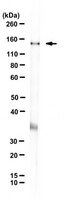
~155 kDa observed; 155.14 kDa calculated. Uncharacterized bands may be observed in some lysate(s).
Physicochemical Information
Dimensions
Materials Information
Toxicological Information
Safety Information according to GHS
Safety Information
Product Usage Statements
Quality Assurance
Evaluated by Western Blotting in HeLa cell lysate.
Western Blotting Analysis: A 1:500 dilution of this antibody detected AKNA in HeLa cell lysate.
Usage Statement
Unless otherwise stated in our catalog or other company documentation accompanying the product(s), our products are intended for research use only and are not to be used for any other purpose, which includes but is not limited to, unauthorized commercial uses, in vitro diagnostic uses, ex vivo or in vivo therapeutic uses or any type of consumption or application to humans or animals.
Storage and Shipping Information
Storage Conditions
Recommend storage at +2°C to +8°C. For long term storage antibodies can be kept at -20°C. Avoid repeated freeze-thaws.