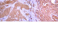
Hypoxia-induced MTA1 promotes MC3T3 osteoblast growth but suppresses MC3T3 osteoblast differentiation.
Liu, T; Zou, W; Shi, G; Xu, J; Zhang, F; Xiao, J; Wang, Y
European journal of medical research
20
10
2015
Mostrar Resumo
Bone fracture is one of the most common physical injuries in which gene expression and the microenvironment are reprogramed to facilitate the recovery process.By specific siRNA transfection and MTT assay, we evaluated the effects of metastasis-associated gene 1 (MTA1) in osteoblast growth. To show the role of MTA1 in osteoblast under hypoxia conditions, by overexpressing and silencing MTA1 expression, we performed mineral deposition and alkaline phosphatase activity assay to observe the differentiation status of osteoblast cells. Real-time PCR and Western blot assays were adopted to detect the expression of certain target genes.Here, we reported that hypoxia-induced MTA1 expression through hypoxia-induced factor 1 alpha (HIF-1α) and stimulated the growth of osteoblast MC3T3 cells. Silencing of MTA1 through specific siRNA suppressed MC3T3 cell growth and elicited cell differentiation and induced alkaline phosphatase activation and the upregulation of bone morphogenetic protein-2 and osteocalcin.We found that MTA1 was regulated by HIF-1α in hypoxia circumstance to suppress osteoblast differentiation. These findings provide new insights for bone fracture healing and new strategies to develop potential targets to promote fracture healing. | Western Blotting | 25644400
 |
Low-dose X-ray irradiation promotes osteoblast proliferation, differentiation and fracture healing.
Chen, M; Huang, Q; Xu, W; She, C; Xie, ZG; Mao, YT; Dong, QR; Ling, M
PloS one
9
e104016
2014
Mostrar Resumo
Great controversy exists regarding the biologic responses of osteoblasts to X-ray irradiation, and the mechanisms are poorly understood. In this study, the biological effects of low-dose radiation on stimulating osteoblast proliferation, differentiation and fracture healing were identified using in vitro cell culture and in vivo animal studies. First, low-dose (0.5 Gy) X-ray irradiation induced the cell viability and proliferation of MC3T3-E1 cells. However, high-dose (5 Gy) X-ray irradiation inhibited the viability and proliferation of osteoblasts. In addition, dynamic variations in osteoblast differentiation markers, including type I collagen, alkaline phosphatase, Runx2, Osterix and osteocalcin, were observed after both low-dose and high-dose irradiation by Western blot analysis. Second, fracture healing was evaluated via histology and gene expression after single-dose X-ray irradiation, and low-dose X-ray irradiation accelerates fracture healing of closed femoral fractures in rats. In low-dose X-ray irradiated fractures, an increase in proliferating cell nuclear antigen (PCNA)-positive cells, cartilage formation and fracture calluses was observed. In addition, we observed more rapid completion of endochondral and intramembranous ossification, which was accompanied by altered expression of genes involved in bone remodeling and fracture callus mineralization. Although the expression level of several osteoblast differentiation genes was increased in the fracture calluses of high-dose irradiated rats, the callus formation and fracture union were delayed compared with the control and low-dose irradiated fractures. These results reveal beneficial effects of low-dose irradiation, including the stimulation of osteoblast proliferation, differentiation and fracture healing, and highlight its potential translational application in novel therapies against bone-related diseases. | Western Blotting | 25089831
 |
Synergistic effects of orbital shear stress on in vitro growth and osteogenic differentiation of human alveolar bone-derived mesenchymal stem cells.
Lim, KT; Hexiu, J; Kim, J; Seonwoo, H; Choung, PH; Chung, JH
BioMed research international
2014
316803
2014
Mostrar Resumo
Cellular behavior is dependent on a variety of physical cues required for normal tissue function. In order to mimic native tissue environments, human alveolar bone-derived mesenchymal stem cells (hABMSCs) were exposed to orbital shear stress (OSS) in a low-speed orbital shaker. The synergistic effects of OSS on proliferation and differentiation of hABMSCs were investigated. In particular, we induced the osteoblastic differentiation of hABMSCs cultured in the absence of OM by exposing hABMSCs to OSS (0.86-1.51 dyne/cm(2)). Activation of Cx43 was associated with exposure of hABMSCs to OSS. The viability of cells stimulated for 10, 30, 60, 120, and 180 min/day increased by approximately 10% compared with that of control. The OSS groups with stimulation of 10, 30, and 60 min/day had more intense mineralized nodules compared with the control group. In quantification of vascular endothelial growth factor (VEGF) and bone morphogenetic protein-2 (BMP-2) protein, VEGF protein levels under stimulation for 10, 60, and 180 min/day and BMP-2 levels under stimulation for 60, 120, and 180 min/day were significantly different compared with those of the control. In conclusion, the results indicated that exposing hABMSCs to OSS enhanced their differentiation and maturation. | | 24575406
 |
Polycystin-1 mediates mechanical strain-induced osteoblastic mechanoresponses via potentiation of intracellular calcium and Akt/β-catenin pathway.
Wang, H; Sun, W; Ma, J; Pan, Y; Wang, L; Zhang, W
PloS one
9
e91730
2014
Mostrar Resumo
Mechanical regulation of bone formation involves a complex biophysical process, yet the underlying mechanisms remain poorly understood. Polycystin-1 (PC1) is postulated to function as a mechanosensory molecule mediating mechanical signal transduction in renal epithelial cells. To investigate the involvement of PC1 in mechanical strain-induced signaling cascades controlling osteogenesis, PKD1 gene was stably silenced in osteoblastic cell line MC3T3-E1 by using lentivirus-mediated shRNA technology. Here, our findings showed that mechanical tensile strain sufficiently enhanced osteogenic gene expressions and osteoblastic proliferation. However, PC1 deficiency resulted in the loss of the ability to sense external mechanical stimuli thereby promoting osteoblastic osteogenesis and proliferation. The signal pathways implicated in this process were intracellular calcium and Akt/β-catenin pathway. The basal levels of intracellular calcium, phospho-Akt, phospho-GSK-3β and nuclear accumulation of active β-catenin were significantly attenuated in PC1 deficient osteoblasts. In addition, PC1 deficiency impaired mechanical strain-induced potentiation of intracellular calcium, and activation of Akt-dependent and Wnt/β-catenin pathways, which was able to be partially reversed by calcium ionophore A23187 treatment. Furthermore, applications of LiCl or A23187 in PC1 deficient osteoblasts could promote osteoblastic differentiation and proliferation under mechanical strain conditions. Therefore, our results demonstrated that osteoblasts require mechanosensory molecule PC1 to adapt to external mechanical tensile strain thereby inducing osteoblastic mechanoresponse, partially through the potentiation of intracellular calcium and downstream Akt/β-catenin signaling pathway. | | 24618832
 |
Effects of electromagnetic fields on osteogenesis of human alveolar bone-derived mesenchymal stem cells.
Lim, K; Hexiu, J; Kim, J; Seonwoo, H; Cho, WJ; Choung, PH; Chung, JH
BioMed research international
2013
296019
2013
Mostrar Resumo
This study was performed to investigate the effects of extremely low frequency pulsed electromagnetic fields (ELF-PEMFs) on the proliferation and differentiation of human alveolar bone-derived mesenchymal stem cells (hABMSCs). Osteogenesis is a complex series of events involving the differentiation of mesenchymal stem cells to generate new bone. In this study, we examined not merely the effect of ELF-PEMFs on cell proliferation, alkaline phosphatase (ALP) activity, and mineralization of the extracellular matrix but vinculin, vimentin, and calmodulin (CaM) expressions in hABMSCs during osteogenic differentiation. Exposure of hABMSCs to ELF-PEMFs increased proliferation by 15% compared to untreated cells at day 5. In addition, exposure to ELF-PEMFs significantly increased ALP expression during the early stages of osteogenesis and substantially enhanced mineralization near the midpoint of osteogenesis within 2 weeks. ELF-PEMFs also increased vinculin, vimentin, and CaM expressions, compared to control. In particular, CaM indicated that ELF-PEMFs significantly altered the expression of osteogenesis-related genes. The results indicated that ELF-PEMFs could enhance early cell proliferation in hABMSCs-mediated osteogenesis and accelerate the osteogenesis. | | 23862141
 |
Inhibition of Rac and ROCK signalling influence osteoblast adhesion, differentiation and mineralization on titanium topographies.
Prowse, PD; Elliott, CG; Hutter, J; Hamilton, DW
PloS one
8
e58898
2013
Mostrar Resumo
Reducing the time required for initial integration of bone-contacting implants with host tissues would be of great clinical significance. Changes in osteoblast adhesion formation and reorganization of the F-actin cytoskeleton in response to altered topography are known to be upstream of osteoblast differentiation, and these processes are regulated by the Rho GTPases. Rac and RhoA (through Rho Kinase (ROCK)). Using pharmacological inhibitors, we tested how inhibition of Rac and ROCK influenced osteoblast adhesion, differentiation and mineralization on PT (Pre-treated) and SLA (sandblasted large grit, acid etched) topographies. Inhibition of ROCK, but not Rac, significantly reduced adhesion number and size on PT, with adhesion size consistent with focal complexes. After 1 day, ROCK, but not Rac inhibition increased osteocalcin mRNA levels on SLA and PT, with levels further increasing at 7 days post seeding. ROCK inhibition also significantly increased bone sialoprotein expression at 7 days, but not BMP-2 levels. Rac inhibition significantly reduced BMP-2 mRNA levels. ROCK inhibition increased nuclear translocation of Runx2 independent of surface roughness. Mineralization of osteoblast cultures was greater on SLA than on PT, but was increased by ROCK inhibition and attenuated by Rac inhibition on both topographies. In conclusion, inhibition of ROCK signalling significantly increases osteoblast differentiation and biomineralization in a topographic dependent manner, and its pharmacological inhibition could represent a new therapeutic to speed bone formation around implanted metals and in regenerative medicine applications. | | 23505566
 |
In vitro effects of low-intensity pulsed ultrasound stimulation on the osteogenic differentiation of human alveolar bone-derived mesenchymal stem cells for tooth tissue engineering.
Lim, K; Kim, J; Seonwoo, H; Park, SH; Choung, PH; Chung, JH
BioMed research international
2013
269724
2013
Mostrar Resumo
Ultrasound stimulation produces significant multifunctional effects that are directly relevant to alveolar bone formation, which is necessary for periodontal healing and regeneration. We focused to find out effects of specific duty cycles and the percentage of time that ultrasound is being generated over one on/off pulse period, under ultrasound stimulation. Low-intensity pulsed ultrasound ((LIPUS) 1 MHz) with duty cycles of 20% and 50% was used in this study, and human alveolar bone-derived mesenchymal stem cells (hABMSCs) were treated with an intensity of 50 mW/cm(2) and exposure time of 10 min/day. hABMSCs exposed at duty cycles of 20% and 50% had similar cell viability (O.D.), which was higher (*P less than 0.05) than that of control cells. The alkaline phosphatase (ALP) was significantly enhanced at 1 week with LIPUS treatment in osteogenic cultures as compared to control. Gene expressions showed significantly higher expression levels of CD29, CD44, COL1, and OCN in the hABMSCs under LIPUS treatment when compared to control after two weeks of treatment. The effects were partially controlled by LIPUS treatment, indicating that modulation of osteogenesis in hABMSCs was related to the specific stimulation. Furthermore, mineralized nodule formation was markedly increased after LIPUS treatment than that seen in untreated cells. Through simple staining methods such as Alizarin red and von Kossa staining, calcium deposits generated their highest levels at about 3 weeks. These results suggest that LIPUS could enhance the cell viability and osteogenic differentiation of hABMSCs, and could be part of effective treatment methods for clinical applications. | Immunofluorescence | 24195067
 |
Lineage differentiation of mesenchymal stem cells from dental pulp, apical papilla, and periodontal ligament.
Kentaro Akiyama,Chider Chen,Stan Gronthos,Songtao Shi
Methods in molecular biology (Clifton, N.J.)
887
2012
Mostrar Resumo
Recently, a variety of mesenchymal stem cells (MSCs), including dental pulp stem cells, stem cells from human exfoliated deciduous teeth, stem cells from apical papilla, periodontal ligament stem cells, and mesenchymal stem cells derived from human gingival, were isolated from orofacial and dental tissues. However, it is unknown whether these orofacial stem cells are derived from mesoderm or neural crest cell. In order to encourage orofacial MSC investigation, we provide detailed protocols for assessing lineage -differentiation of orofacial MSCs. | | 22566051
 |
In vitro and in vivo characteristics of stem cells derived from the periodontal ligament of human deciduous and permanent teeth.
Je Seon Song,Seong-Oh Kim,Seung-Hye Kim,Hyung-Jun Choi,Heung-Kyu Son,Han-Sung Jung,Chang-Sung Kim,Jae-Ho Lee
Tissue engineering. Part A
18
2012
Mostrar Resumo
In many studies, adult stem cells have been found in human periodontal ligament (PDL), but in most cases they were found in the permanent teeth. The aim of the present study was to characterize stem cells from the PDL of deciduous teeth (dPDLSCs) and compare them with those from the PDL of permanent teeth (pPDLSCs). Stem cell markers were examined by a flow cytometric analysis. The results of in vitro differentiation into adipogenic and osteogenic lineages were analyzed by histochemical staining and quantitative reverse transcription-polymerase chain reaction (RT-PCR). The results of in vivo transplantation were analyzed by histological staining, immunohistochemical staining, and quantitative RT-PCR. There were no significant differences in the proliferation rate, cell cycle distribution, expressions of stem cell markers such as Stro-1 and CD146, or in vitro differentiation. The pPDLSC transplants made more typical cementum/PDL-like tissues and expressed more cementum/PDL-related genes (CP23 and collagen XII) than did the dPDLSC transplants. Together, these results suggest that pPDLSCs are better candidates for use in reconstructing periodontium. | | 22571499
 |


