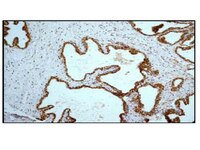
Astrocytic adenosine receptor A2A and Gs-coupled signaling regulate memory.
Orr, AG; Hsiao, EC; Wang, MM; Ho, K; Kim, DH; Wang, X; Guo, W; Kang, J; Yu, GQ; Adame, A; Devidze, N; Dubal, DB; Masliah, E; Conklin, BR; Mucke, L
Nature neuroscience
18
423-34
2015
Mostrar Resumo
Astrocytes express a variety of G protein-coupled receptors and might influence cognitive functions, such as learning and memory. However, the roles of astrocytic Gs-coupled receptors in cognitive function are not known. We found that humans with Alzheimer's disease (AD) had increased levels of the Gs-coupled adenosine receptor A2A in astrocytes. Conditional genetic removal of these receptors enhanced long-term memory in young and aging mice and increased the levels of Arc (also known as Arg3.1), an immediate-early gene that is required for long-term memory. Chemogenetic activation of astrocytic Gs-coupled signaling reduced long-term memory in mice without affecting learning. Like humans with AD, aging mice expressing human amyloid precursor protein (hAPP) showed increased levels of astrocytic A2A receptors. Conditional genetic removal of these receptors enhanced memory in aging hAPP mice. Together, these findings establish a regulatory role for astrocytic Gs-coupled receptors in memory and suggest that AD-linked increases in astrocytic A2A receptor levels contribute to memory loss. | 25622143
 |
Re-evaluation of the role of calcium homeostasis endoplasmic reticulum protein (CHERP) in cellular calcium signaling.
Lin-Moshier, Y; Sebastian, PJ; Higgins, L; Sampson, ND; Hewitt, JE; Marchant, JS
The Journal of biological chemistry
288
355-67
2013
Mostrar Resumo
Changes in cytoplasmic Ca(2+) concentration, resulting from activation of intracellular Ca(2+) channels within the endoplasmic reticulum, regulate several aspects of cellular growth and differentiation. Ca(2+) homeostasis endoplasmic reticulum protein (CHERP) is a ubiquitously expressed protein that has been proposed as a regulator of both major families of endoplasmic reticulum Ca(2+) channels, inositol 1,4,5-trisphosphate receptors (IP(3)Rs) and ryanodine receptors (RyRs), with resulting effects on mitotic cycling. However, the manner by which CHERP regulates intracellular Ca(2+) channels to impact cellular growth is unknown. Here, we challenge previous findings that CHERP acts as a direct cytoplasmic regulator of IP(3)Rs and RyRs and propose that CHERP acts in the nucleus to impact cellular proliferation by regulating the function of the U2 snRNA spliceosomal complex. The previously reported effects of CHERP on cellular growth therefore are likely indirect effects of altered spliceosomal function, consistent with prior data showing that loss of function of U2 snRNP components can interfere with cell growth and induce cell cycle arrest. | 23148228
 |
Increasing CREB function in the CA1 region of dorsal hippocampus rescues the spatial memory deficits in a mouse model of Alzheimer's disease.
Yiu, AP; Rashid, AJ; Josselyn, SA
Neuropsychopharmacology : official publication of the American College of Neuropsychopharmacology
36
2169-86
2011
Mostrar Resumo
The principal defining feature of Alzheimer's disease (AD) is memory impairment. As the transcription factor CREB (cAMP/Ca(2+) responsive element-binding protein) is critical for memory formation across species, we investigated the role of CREB in a mouse model of AD. We found that TgCRND8 mice exhibit a profound impairment in the ability to form a spatial memory, a process that critically relies on the dorsal hippocampus. Perhaps contributing to this memory deficit, we observed additional deficits in the dorsal hippocampus of TgCRND8 mice in terms of (1) biochemistry (decreased CREB activation in the CA1 region), (2) neuronal structure (decreased spine density and dendritic complexity of CA1 pyramidal neurons), and (3) neuronal network activity (decreased arc mRNA levels following behavioral training). Locally and acutely increasing CREB function in the CA1 region of dorsal hippocampus of TgCRND8 mice was sufficient to restore function in each of these key domains (biochemistry, neuronal structure, network activity, and most importantly, memory formation). The rescue produced by increasing CREB was specific both anatomically and behaviorally and independent of plaque load or Aβ levels. Interestingly, humans with AD show poor spatial memory/navigation and AD brains have disrupted (1) CREB activation, and (2) spine density and dendritic complexity in hippocampal CA1 pyramidal neurons. These parallel findings not only confirm that TgCRND8 mice accurately model key aspects of human AD, but furthermore, suggest the intriguing possibility that targeting CREB may be a useful therapeutic strategy in treating humans with AD. | 21734652
 |
Identification of drug candidates which increase cytochrome c oxidase activity in deficient patient fibroblasts.
Maj M, Sriskandarajah N, Hung V, Browne I, Shah B, Weadge A, Jamieson NL, Tropak M, Cameron JM, Addis JB, Robinson BH
Mitochondrion
2010
Mostrar Resumo
Cytochrome c oxidase (COX) activity reflects the expressed level of respiratory chain complexes, mtDNA levels, titer and mass of mitochondria. Activity is also indicative of the overall fitness of mt-transcription factors and the import, transcription and translation of mt-proteins. We have developed a high-throughput assay to measure COX activity using live cells to screen chemical libraries for compounds capable of increasing COX activity. These libraries have revealed four examples which elevated the activities of COX in NIH-3T3 fibroblasts and in fibroblasts from patients with COX defects independent of the peroxisome proliferator activated receptor family. | 21050896
 |
Stimulated neuronal expression of brain-derived neurotrophic factor by Neurotropin.
Yu Fukuda,Thomas L Berry,Matthew Nelson,Christopher L Hunter,Koki Fukuhara,Hideki Imai,Shinji Ito,Ann-Charlotte Granholm-Bentley,Allen P Kaplan,Tatsuro Mutoh
Molecular and cellular neurosciences
45
2010
Mostrar Resumo
Expression of brain-derived neurotrophic factor (BDNF) was stimulated in human neuroblastoma SH-SY5Y cells by a nonprotein extract of inflamed rabbit skin inoculated with vaccinia virus (Neurotropin), an analgesic widely used in Japan for treatment of disorders associated with chronic pain, with the optimal dosage at 10mNU/mL. This stimulation was accompanied by activations of p42/44 MAP kinase, CREB and c-Fos expression. Inhibitors of MAP kinases or PI 3-kinase prevented the stimulatory action of Neurotropin, indicating that neuronal TrkB/CREB pathway mediates the action. Repetitive oral administration of Neurotropin (200NU/kg/day, 3months) prevented the age-dependent decline in hippocampal BDNF expression in Ts65Dn mice, a model of Down's syndrome. This effect was associated with the improvement of spatial cognition of the mice. These results open an intriguing new strategy in which Neurotropin may prove beneficial treatment for neurodegenerative disorders. | 20600926
 |


