Millipore Sigma Vibrant Logo
Attention: We have moved. Merck Millipore products are no longer available for purchase on MerckMillipore.com.Learn More

324867 PhosphoDetect™ Anti-EGFR (pTyr¹⁰⁶⁸) Rabbit pAb

324867
Purchase on Sigma-Aldrich

Aperçu

Replacement Information

Tableau de caractéristiques principal

Species ReactivityHostAntibody Type
Ch, H, M, RRbPolyclonal Antibody

Products

RéférenceConditionnement Qté
324867-10T Ampoule plast. 10 t
Description
OverviewRecognizes the ~185 kDa EGFR protein phosphorylated at Tyr1068. Note: 1 T = 1 test. Sufficient for 10 Western miniblots.
Catalogue Number324867
Brand Family Calbiochem®
SynonymsAnti-Epidermal Growth Factor Receptor (pTyr¹⁰⁶⁸)
Application Data
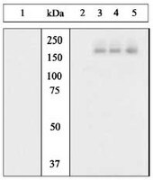
Detection of human EGFR, phosphorylated at Tyr1068, by immunoblotting. Samples: Whole cell lysate from A431 cells unstimulated (lane 1), or stimulated for EGF (lanes 2-5). Primary antibody: PhosphoDetect™ Anti-Epidermal Growth Factor Receptor (pTyr1068) Rabbit pAb (Cat. No. 324867) (1:1000) pre-incubated without peptide (lanes 1 and 5), with phosphopeptide immunogen (lane 2), a generic phosphotyrosine-containing peptide (lane 3), or non-phosphopeptide corresponding to the immunogen (lane 4). Detection: chemiluminescence.
References
ReferencesJo, M., et al. 2000. J. Biol. Chem. 275, 8806.
Maudsley, S., et al. 2000. J. Biol. Chem. 275, 9572.
Sieg, D.J. et al. 2000. Nat. Cell. Biol. 2, 249.
Barbier, A.J. et al. 1999. J. Biol. Chem. 274, 14067.
Hashimoto, A. et al. 1999. J. Biol. Chem. 274, 20139
Poppleton, H.M. et al. 1999. Arch. Biochem. Biophys. 363, 227.
Batzer, A.G. et al. 1994. Mol. Cell. Biol. 14, 5192.
Product Information
FormLiquid
FormulationIn Dulbecco's PBS, 1 mg/ml BSA, 50% glycerol, pH 7.4.
Preservative≤0.1% sodium azide
Quality LevelMQ100
Applications
Key Applications Immunoblotting (Western Blotting)
Application NotesImmunoblotting (1:1000)
Application CommentsWhen diluted 1:1000 there is sufficient antibody to perform 10 miniblots (10 T) at 10 ml per blot. Variables associated with assay conditions will dictate the proper working dilution.
Biological Information
Immunogena synthetic phosphopeptide corresponding to amino acids surrounding the Tyr¹⁰⁶⁸ phosphorylation site of human EGFR
ImmunogenHuman
HostRabbit
IsotypeIgG
Species Reactivity
  • Chicken
  • Human
  • Mouse
  • Rat
Antibody TypePolyclonal Antibody
Physicochemical Information
Dimensions
Materials Information
Toxicological Information
Safety Information according to GHS
Safety Information
Product Usage Statements
Storage and Shipping Information
Ship Code Dry Ice Only
Toxicity Standard Handling
Storage -20°C
Avoid freeze/thaw Avoid freeze/thaw
Do not freeze Ok to freeze
Special InstructionsFollowing initial thaw, aliquot and freeze (-70°C).
Packaging Information
Transport Information
Supplemental Information
Specifications
Global Trade Item Number
Référence GTIN
324867-10T 04055977215823

Documentation

PhosphoDetect™ Anti-EGFR (pTyr¹⁰⁶⁸) Rabbit pAb FDS

Titre

Fiche de données de sécurité des matériaux (FDS) 

PhosphoDetect™ Anti-EGFR (pTyr¹⁰⁶⁸) Rabbit pAb Certificats d'analyse

TitreNuméro de lot
324867

Références bibliographiques

Aperçu de la référence bibliographique
Jo, M., et al. 2000. J. Biol. Chem. 275, 8806.
Maudsley, S., et al. 2000. J. Biol. Chem. 275, 9572.
Sieg, D.J. et al. 2000. Nat. Cell. Biol. 2, 249.
Barbier, A.J. et al. 1999. J. Biol. Chem. 274, 14067.
Hashimoto, A. et al. 1999. J. Biol. Chem. 274, 20139
Poppleton, H.M. et al. 1999. Arch. Biochem. Biophys. 363, 227.
Batzer, A.G. et al. 1994. Mol. Cell. Biol. 14, 5192.
Fiche technique

Note that this data sheet is not lot-specific and is representative of the current specifications for this product. Please consult the vial label and the certificate of analysis for information on specific lots. Also note that shipping conditions may differ from storage conditions.

Revision03-March-2008 JSW
SynonymsAnti-Epidermal Growth Factor Receptor (pTyr¹⁰⁶⁸)
ApplicationImmunoblotting (1:1000)
Application Data
Detection of human EGFR, phosphorylated at Tyr1068, by immunoblotting. Samples: Whole cell lysate from A431 cells unstimulated (lane 1), or stimulated for EGF (lanes 2-5). Primary antibody: PhosphoDetect™ Anti-Epidermal Growth Factor Receptor (pTyr1068) Rabbit pAb (Cat. No. 324867) (1:1000) pre-incubated without peptide (lanes 1 and 5), with phosphopeptide immunogen (lane 2), a generic phosphotyrosine-containing peptide (lane 3), or non-phosphopeptide corresponding to the immunogen (lane 4). Detection: chemiluminescence.
DescriptionRabbit polyclonal antibody adsorbed against non-phosphorylation site, followed by immunoaffinity chromatography. Recognizes the ~170 kDa EGFR protein phosphorylated at Tyr1068.
BackgroundEpidermal Growth Factor (EGF) is responsible for a variety of biological effects including mitogenesis and differentiation. The EGF receptor (EGFR) has three domains: the extracellular, transmembrane, and cytoplasmic domains. Upon binding of EGF to the extracellular domain, dimerization of the receptor occurs and the cytoplasmic domains display tyrosine kinnase activity. This leads to autophosphorylation of the ~170 kDa glycoprotein on various residues within the cytoplasmic domain as well as phosphorylation of intracellular substrates. Tyr1068 is an autophosphorylation site in EGFR. The SH2 domain of Grb2 binds to Tyr1068 of EGFR in the multimeric complex EGFR-Grb2-mSos1 that is formed upon stimulation of EGFR with EGF.
HostRabbit
Immunogen speciesHuman
Immunogena synthetic phosphopeptide corresponding to amino acids surrounding the Tyr¹⁰⁶⁸ phosphorylation site of human EGFR
IsotypeIgG
Specieschicken, human, mouse, rat
FormLiquid
FormulationIn Dulbecco's PBS, 1 mg/ml BSA, 50% glycerol, pH 7.4.
Preservative≤0.1% sodium azide
CommentsWhen diluted 1:1000 there is sufficient antibody to perform 10 miniblots (10 T) at 10 ml per blot. Variables associated with assay conditions will dictate the proper working dilution.
Storage Avoid freeze/thaw
-20°C
Do Not Freeze Ok to freeze
Special InstructionsFollowing initial thaw, aliquot and freeze (-70°C).
Toxicity Standard Handling
ReferencesJo, M., et al. 2000. J. Biol. Chem. 275, 8806.
Maudsley, S., et al. 2000. J. Biol. Chem. 275, 9572.
Sieg, D.J. et al. 2000. Nat. Cell. Biol. 2, 249.
Barbier, A.J. et al. 1999. J. Biol. Chem. 274, 14067.
Hashimoto, A. et al. 1999. J. Biol. Chem. 274, 20139
Poppleton, H.M. et al. 1999. Arch. Biochem. Biophys. 363, 227.
Batzer, A.G. et al. 1994. Mol. Cell. Biol. 14, 5192.