Millipore Sigma Vibrant Logo
Attention: We have moved. Merck Millipore products are no longer available for purchase on MerckMillipore.com.Learn More

ABE1868 Anti-TDRD3

ABE1868
100 µL  
Purchase on Sigma-Aldrich

Aperçu

Replacement Information

Tableau de caractéristiques principal

Species ReactivityKey ApplicationsHostFormatAntibody Type
HIP, ChIP, WBRbUnpurifiedPolyclonal Antibody
Description
Catalogue NumberABE1868
DescriptionAnti-TDRD3
Alternate Names
  • Tudor domain-containing protein 3
Background InformationTudor domain-containing protein 3 (UniProt: Q9H7E2; also known as TDRD3) is encoded by the TDRD3 gene (Gene ID: 81550) in human. TDRD3 belongs to an evolutionarily conserved family of 12 proteins (TDRD1-TDRD12) that contain one or more Tudor domains, which recognize symmetric dimethylarginine (sDMA) residues with the exception of TDRD3, which associates selectively with asymmetric dimethylarginine. TDRD3, in addition to its Tudor domain, contains a putative nucleic acid recognition motif and an ubiquitin-associated domain. Tudor domains are highly conserved throughout evolution: they can be found in virtually all organisms, from bacteria to mammals. TDRD3 has been reported in heart, brain, placenta, lung, liver, skeletal muscle, kidney, and pancreas. It is found both in the nucleus and in cytoplasm. In nucleus, it acts as a coactivator and recognizes and binds asymmetric dimethylation on the core histone tails. The Tudor domain specifically recognizes and binds asymmetric dimethylation of histone H3 'Arg-17' (H3R17me2a) and histones H4 'Arg-3', 2 tags for epigenetic transcriptional activation. In cytoplasm, it may play a role in the assembly and/or disassembly of mRNA stress granules and in the regulation of translation of target mRNAs by binding Arg/Gly-rich motifs (GAR) in dimethylarginine-containing proteins. TDRD3 is reported to act as a positive regulator of c-MYC gene transcription by recruiting topoisomerase IIIB (TOP3B) to the c-MYC gene promoter region via H4R3me2a recognition. Overexpression of TDRD3 gene has a strong predictive value for poor prognosis of estrogen receptor-negative breast cancers. (Ref.: Goulet, I et al. (2008). Hum. Mol. Genet. 17(19):3055-3074; Sikorsky, T et al. (2012). Nucleic Acids Res. 40(22):11748-55).
References
Product Information
FormatUnpurified
PresentationRabbit polyclonal antiserum with 0.05% sodium azide.
Quality LevelMQ100
Applications
ApplicationAnti-TDRD3, Cat. No. ABE1868, is a highly specific rabbit polyclonal antibody that targets Tudor domain-containing protein 3 and has been tested in Chromatin Immunoprecipitation (ChIP), Immunoprecipitation, and Western Blotting.
Key Applications
  • Immunoprecipitation
  • Chromatin Immunoprecipitation (ChIP)
  • Western Blotting
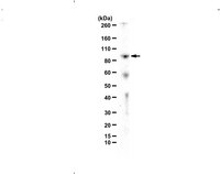
Application NotesWestern Blotting Analysis: A 1:500 dilution from a representative lot detected TDRD3 in 20 µL of MCF7 cell lysate.

Immunoprecipitation Analysis: A representative lot detected TDRD3 in Immunoprecipitation applications (Yang, Y., et. al. (2014). Mol Cell. 53(3):484-97).

Chromatin Immunoprecipitation (ChIP) Analysis: A representative lot detected TDRD3 in Chromatin Immunoprecipitation applications (Yang, Y., et. al. (2014). Mol Cell. 53(3):484-97;Yang, Y., et. al. (2010). Mol Cell. 40(6):1016-23).
Biological Information
ImmunogenGST-tagged recombinant fragment corresponding to 258 amino acids from the C-terminal half of human Tudor domain-containing protein 3.
Epitopedomain of:
ConcentrationPlease refer to lot specific datasheet.
HostRabbit
SpecificityThis polyclonal antibody detects the Tudor domain containing protein 3 in human. It targets an epitope within 258 amino acids in the C-terminal half.
IsotypeIgG
Species Reactivity
  • Human
Species Reactivity NoteHuman.
Antibody TypePolyclonal Antibody
Entrez Gene Number
Gene Symbol
  • TDRD3
Purification MethodUnpurified
UniProt Number
Molecular Weight~85 kDa observed; 73.18 kDa calculated. Uncharacterized bands may be observed in some lysate(s).
Physicochemical Information
Dimensions
Materials Information
Toxicological Information
Safety Information according to GHS
Safety Information
Product Usage Statements
Quality AssuranceEvaluated by Immunoprecipitation in MCF7 cell lysate.

Immunoprecipitation Analysis: 10 µg of this antibody immunoprecipitated TDRD3 in 500 µg of MCF7 cell lysate.
Usage Statement
  • Unless otherwise stated in our catalog or other company documentation accompanying the product(s), our products are intended for research use only and are not to be used for any other purpose, which includes but is not limited to, unauthorized commercial uses, in vitro diagnostic uses, ex vivo or in vivo therapeutic uses or any type of consumption or application to humans or animals.
Storage and Shipping Information
Storage ConditionsStable for 1 year at -20°C from date of receipt.

Handling Recommendations: Upon receipt and prior to removing the cap, centrifuge the vial and gently mix the solution. Aliquot into microcentrifuge tubes and store at -20°C. Avoid repeated freeze/thaw cycles, which may damage IgG and affect product performance.
Packaging Information
Material Size100 µL
Transport Information
Supplemental Information
Specifications
Global Trade Item Number
Référence GTIN
ABE1868 04054839178986