Survey for human polyomaviruses in cancer.
Toptan, T; Yousem, SA; Ho, J; Matsushima, Y; Stabile, LP; Fernández-Figueras, MT; Bhargava, R; Ryo, A; Moore, PS; Chang, Y
JCI Insight
1
2015
Afficher le résumé
Over the past 8 years, the discovery of 11 new human polyomaviruses (HPyVs) has revived interest in this DNA tumor virus family. Although HPyV infection is widespread and largely asymptomatic, one of these HPyVs, Merkel cell polyomavirus (MCV), is a bona fide human tumor virus. JC virus (JCV), BK virus, HPyV7, and trichodysplasia-spinulosa virus (TSV) can cause nonneoplastic diseases in the setting of immunosuppression. Few specific reagents are available to study the biology of the newly discovered HPyVs. We developed a pan-HPyV-screening method using a cocktail of 3 antibodies that, when combined, recognize T antigen proteins of all HPyVs. We validated detection characteristics of the antibody cocktail by immunoblotting and immunohistochemistry and screened 1,184 cases, including well-defined diseases and tumor tissue microarrays. This assay robustly detected MCV, TSV, JCV, and HPyV7 in etiologically related diseases. We further identified WU polyomavirus in a case of chronic lymphocytic lymphoma-associated bronchitis. Except for scattered, incidentally infected cells in 5% of lung squamous cell carcinomas and colon adenocarcinomas, a broad panel of tumor tissues was largely negative for infection by any HPyV. This method eliminates known HPyVs as suspected causes of cancers investigated in this study. Pan-HPyV survey can be applied to identify diseases associated with recently discovered polyomaviruses. | 27034991
 |
Bromodomain protein Brd4 plays a key role in Merkel cell polyomavirus DNA replication.
Wang, X; Li, J; Schowalter, RM; Jiao, J; Buck, CB; You, J
PLoS Pathog
8
e1003021
2011
Afficher le résumé
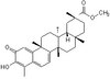
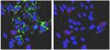
Merkel cell polyomavirus (MCV or MCPyV) is the first human polyomavirus to be definitively linked to cancer. The mechanisms of MCV-induced oncogenesis and much of MCV biology are largely unexplored. In this study, we demonstrate that bromodomain protein 4 (Brd4) interacts with MCV large T antigen (LT) and plays a critical role in viral DNA replication. Brd4 knockdown inhibits MCV replication, which can be rescued by recombinant Brd4. Brd4 colocalizes with the MCV LT/replication origin complex in the nucleus and recruits replication factor C (RFC) to the viral replication sites. A dominant negative inhibitor of the Brd4-MCV LT interaction can dissociate Brd4 and RFC from the viral replication complex and abrogate MCV replication. Furthermore, obstructing the physiologic interaction between Brd4 and host chromatin with the chemical compound JQ1(+) leads to enhanced MCV DNA replication, demonstrating that the role of Brd4 in MCV replication is distinct from its role in chromatin-associated transcriptional regulation. Our findings demonstrate mechanistic details of the MCV replication machinery; providing novel insight to elucidate the life cycle of this newly discovered oncogenic DNA virus. | 23144621
|