Millipore Sigma Vibrant Logo
Attention: We have moved. Merck Millipore products are no longer available for purchase on MerckMillipore.com.Learn More

MABE952 Anti-Histone H3.1 Antibody, clone 1D4F2

MABE952
100 µg  
Purchase on Sigma-Aldrich

Offres spéciales

Aperçu

Replacement Information

Offres spéciales

Tableau de caractéristiques principal

Species ReactivityKey ApplicationsHostFormatAntibody Type
H, MWB, ICC, IP, ChIP, ChIP-seqRPurifiedMonoclonal Antibody
Description
Catalogue NumberMABE952
DescriptionAnti-Histone H3.1 Antibody, clone 1D4F2
Alternate Names
  • Histone H3.1
  • Histone H3/a
  • Histone H3/b
  • Histone H3/c
  • Histone H3/d
  • Histone H3/f
  • Histone H3/h
  • Histone H3/i
  • Histone H3/j
  • Histone H3/k
  • Histone H3/l
Background InformationHistone H3 is one of the five main histone proteins involved in the structure of chromatin in eukaryotic cells. Featuring a main globular domain and a long N-terminal tail, H3 is involved with the structure of the nucleosomes of the 'beads on a string' structure. The N-terminal tail of histone H3 protrudes from the globular nucleosome core and can undergo several different types of epigenetic modifications that influence cellular processes. These modifications include the covalent attachment of methyl or acetyl groups to lysine and arginine amino acids and the phosphorylation of serine or threonine.
References
Product Information
FormatPurified
PresentationPurified rat monoclonal in buffer containing 0.1 M Tris-Glycine (pH 7.4), 150 mM NaCl with 0.05% sodium azide.
Quality LevelMQ100
Applications
ApplicationThis Anti-Histone H3.1 Antibody, clone 1D4F2 is validated for use in western blotting, ICC, IP, ChIP & ChIP-seq for the detection of Histone H3.1.
Key Applications
  • Western Blotting
  • Immunocytochemistry
  • Immunoprecipitation
  • Chromatin Immunoprecipitation (ChIP)
  • ChIP-seq
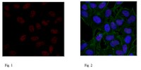
Application NotesImmunocytochemistry Analysis: A 1:50 dilution from a representative lot detected Histone H3.1 in HeLa cells.

Immunocytochemistry Analysis: A 1:2,000 dilution from a representative lot detected Histone H3.1 in NIH/3T3 cells (Prof. Taro Tachibana, Cell Engineering Corporation.).

Alexa Fluor® is a registered trademark of Life Technologies®.

Immunoprecipitation Analysis: A representative lot from an independent laboratory detected Histone H3.1 in C2C12 cells (Harada, A., et al. (2012). EMBO J. 31(13):2994-3007.).

Chromatin Immunoprecipitation Analysis: A representative lot from an independent laboratory immunoprecipitated Histone H3.1 (Maehara, K., et al. (2013). Nucleic Acids Res. 41(1):54-62.).

Chromatin Immunoprecipitation Sequencing Analysis: A representative lot from an independent laboratory immunoprecipitated Histone H3.1 (Maehara, K., et al. (2013). Nucleic Acids Res. 41(1):54-62.).
Biological Information
ImmunogenKLH-conjugated linear peptide corresponding to human Histone H3.1.
Clone1D4F2
Concentration1 mg/mL
HostRat
Species Reactivity
  • Human
  • Mouse
Antibody TypeMonoclonal Antibody
Entrez Gene Number
Gene Symbol
  • HIST1H3A
  • H3FA
  • HIST1H3B
  • H3FL
  • HIST1H3C
  • H3FC
  • HIST1H3D
  • H3FB
  • HIST1H3E
  • H3FD
  • HIST1H3F
  • H3FI
  • HIST1H3G
  • H3FH
  • HIST1H3H
  • H3FK
  • HIST1H3I
  • H3FF
  • HIST1H3J
  • H3FJ
Purification MethodProtein G Purified
UniProt Number
Molecular Weight~17 kDa observed
Physicochemical Information
Dimensions
Materials Information
Toxicological Information
Safety Information according to GHS
Safety Information
Product Usage Statements
Quality AssuranceEvaluated by Western Blotting in HeLa acid extract.

Western Blotting Analysis: 1 µg/mL of this antibody detected Histone H3.1 in 10 µg of HeLa acid extract.
Usage Statement
  • Unless otherwise stated in our catalog or other company documentation accompanying the product(s), our products are intended for research use only and are not to be used for any other purpose, which includes but is not limited to, unauthorized commercial uses, in vitro diagnostic uses, ex vivo or in vivo therapeutic uses or any type of consumption or application to humans or animals.
Storage and Shipping Information
Storage ConditionsStable for 1 year at 2-8°C from date of receipt.
Packaging Information
Material Size100 µg
Transport Information
Supplemental Information
Specifications
Global Trade Item Number
Référence GTIN
MABE952 04053252975691

Documentation

Anti-Histone H3.1 Antibody, clone 1D4F2 FDS

Titre

Fiche de données de sécurité des matériaux (FDS) 

Anti-Histone H3.1 Antibody, clone 1D4F2 Certificats d'analyse

TitreNuméro de lot
Anti-Histone H3.1, clone 1D4F2 - Q2426892 Q2426892
Anti-Histone H3.1, clone 1D4F2 - 3317043 3317043
Anti-Histone H3.1, clone 1D4F2 - 3526337 3526337
Anti-Histone H3.1, clone 1D4F2 - 3888819 3888819
Anti-Histone H3.1, clone 1D4F2 - 3927175 3927175
Anti-Histone H3.1, clone 1D4F2 - 4037958 4037958
Anti-Histone H3.1, clone 1D4F2 -2683301 2683301
Anti-Histone H3.1, clone 1D4F2 Monoclonal Antibody 3095014
Anti-Histone H3.1, clone 1D4F2 Monoclonal Antibody 3033911

Références bibliographiques

Aperçu de la référence bibliographiqueNº PubMed
A co-localization model of paired ChIP-seq data using a large ENCODE data set enables comparison of multiple samples.
Maehara, Kazumitsu, et al.
Nucleic Acids Res., 41: 54-62 (2013)  2013

Afficher le résumé
23125363 23125363

Informations techniques

Titre
White Paper - The Message in the Marks: Deciphering Cancer Epigenetics