Long noncoding RNA EWSAT1-mediated gene repression facilitates Ewing sarcoma oncogenesis.
Marques Howarth, M; Simpson, D; Ngok, SP; Nieves, B; Chen, R; Siprashvili, Z; Vaka, D; Breese, MR; Crompton, BD; Alexe, G; Hawkins, DS; Jacobson, D; Brunner, AL; West, R; Mora, J; Stegmaier, K; Khavari, P; Sweet-Cordero, EA
The Journal of clinical investigation
124
5275-90
2014
Show Abstract
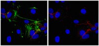
Chromosomal translocation that results in fusion of the genes encoding RNA-binding protein EWS and transcription factor FLI1 (EWS-FLI1) is pathognomonic for Ewing sarcoma. EWS-FLI1 alters gene expression through mechanisms that are not completely understood. We performed RNA sequencing (RNAseq) analysis on primary pediatric human mesenchymal progenitor cells (pMPCs) expressing EWS-FLI1 in order to identify gene targets of this oncoprotein. We determined that long noncoding RNA-277 (Ewing sarcoma-associated transcript 1 [EWSAT1]) is upregulated by EWS-FLI1 in pMPCs. Inhibition of EWSAT1 expression diminished the ability of Ewing sarcoma cell lines to proliferate and form colonies in soft agar, whereas EWSAT1 inhibition had no effect on other cell types tested. Expression of EWS-FLI1 and EWSAT1 repressed gene expression, and a substantial fraction of targets that were repressed by EWS-FLI1 were also repressed by EWSAT1. Analysis of RNAseq data from primary human Ewing sarcoma further supported a role for EWSAT1 in mediating gene repression. We identified heterogeneous nuclear ribonucleoprotein (HNRNPK) as an RNA-binding protein that interacts with EWSAT1 and found a marked overlap in HNRNPK-repressed genes and those repressed by EWS-FLI1 and EWSAT1, suggesting that HNRNPK participates in EWSAT1-mediated gene repression. Together, our data reveal that EWSAT1 is a downstream target of EWS-FLI1 that facilitates the development of Ewing sarcoma via the repression of target genes. | 25401475
 |
Phase II study of Cediranib (AZD 2171), an inhibitor of the vascular endothelial growth factor receptor, for second-line therapy of small cell lung cancer (National Cancer Institute #7097).
Ramalingam, SS; Belani, CP; Mack, PC; Vokes, EE; Longmate, J; Govindan, R; Koczywas, M; Ivy, SP; Gandara, DR
Journal of thoracic oncology : official publication of the International Association for the Study of Lung Cancer
5
1279-84
2010
Show Abstract
Inhibition of angiogenesis is a novel strategy for the treatment of cancer. We evaluated the safety and efficacy of cediranib, a potent small molecule inhibitor of the vascular endothelial growth factor receptor, in patients with refractory or recurrent small cell lung cancer (SCLC).Patients with SCLC with progression after prior platinum-based chemotherapy only; performance status (PS) of 0 to 2; and adequate bone marrow, renal, and hepatic function were included. The dose of cediranib was 45 mg PO once a day for the first 12 patients and was reduced to 30 mg PO once a day for the subsequent patients because of intolerance of the higher dose. Treatment was given on a daily continuous schedule. The primary end point was determination of the response rate.Twenty-five patients were enrolled. Patient characteristics were as follows: 13 men; median age 61 years; PS 0 (12 pts), PS 1 (12 pts). A median of two cycles were administered. Salient grade 3/4 toxicities were fatigue, diarrhea, hypertension, proteinuria, and elevated liver enzymes. Tolerability was better with the 30 mg dose once a day. Nine patients had stable disease, but none had a confirmed partial response. The median progression-free survival and overall survival were 2 and 6 months, respectively. Response criteria to proceed to full accrual were not met. Increase in circulating endothelial cell count was noted at the time of progression in several patients.Cediranib failed to demonstrate objective responses in recurrent or refractory SCLC at the dose and schedule evaluated. The 45 mg dose was intolerable in a majority of SCLC patients. | 20559150
|
The Schneiderian membrane contains osteoprogenitor cells: in vivo and in vitro study.
S Srouji, T Kizhner, D Ben David, M Riminucci, P Bianco, E Livne
Calcified tissue international
84
138-45
2008
Show Abstract
Recent studies successfully demonstrated induction of new bone formation in the maxillary sinus by mucosal membrane lifting without the use of any graft material. The aim of this work was to test the osteogenic potential of human maxillary sinus Schneiderian membrane (hMSSM) using both in vitro and in vivo assays. Samples of hMSSM were used for establishment of cell cultures and for histological studies. Flow cytometry analysis was performed on P(0), P(1), and P(2) cultures using established mesenchymal progenitor cell markers (CD 105, CD 146, CD 71, CD 73, CD 166), and the ability of hMSSM cells to undergo osteogenic differentiation in culture was analyzed using relevant in vitro assays. Results showed that hMSSM cells could be induced to express alkaline phosphatase, bone morphogenic protein-2, osteopontin, osteonectin, and osteocalcin and to mineralize their extracellular matrix. Inherent osteogenic potential of hMSSM-derived cells was further proven by in vivo experiments, which demonstrated the formation of histology-proven bone at ectopic sites following transplantation of hMSSM-derived cells in conjunction with an osteoconductive scaffold. This study provides the biological background for understanding the observed clinical phenomena in sinus lifting. Our results show that a genuine osteogenic potential is associated with the hMSSM and can contribute to development of successful sinus augmentation techniques. | 19067018
|
PLXDC1 (TEM7) is identified in a genome-wide expression screen of glioblastoma endothelium.
Robert M Beaty,Jennifer B Edwards,Kathy Boon,I-Mei Siu,James E Conway,Gregory J Riggins
Journal of neuro-oncology
81
2007
Show Abstract
Glioblastomas are a highly aggressive brain tumor, with one of the highest rates of new blood vessel formation. In this study we used a combined experimental and bioinformatics strategy to determine which genes were highly expressed and specific for glioblastoma endothelial cells (GBM-ECs), compared to gene expression in normal tissue and endothelium. Starting from fresh glioblastomas, several rounds of negative and positive selection were used to isolate GBM-ECs and extract total RNA. Using Serial Analysis of Gene Expression (SAGE), 116,259 transcript tags (35,833 unique tags) were sequenced. From this expression analysis, we found 87 tags that were not expressed in normal brain. Further subtraction of normal endothelium, bone marrow, white blood cell and other normal tissue transcripts resulted in just three gene transcripts, ANAPC10, PLXDC1(TEM7), and CYP27B1, that are highly specific to GBM-ECs. Immunohistochemistry with an antibody for PLXDC1 showed protein expression in GBM microvasculature, but not in the normal brain endothelium tested. Our results suggest that this study succeeded in identifying GBM-EC specific genes. The entire gene expression profile for the GBM-ECs and other tissues used in this study are available at SAGE Genie (http://cgap.nci.nih.gov/SAGE). Functionally, the protein products of the three tags most specific to GBM-ECs have been implicated in processes critical to endothelial cell proliferation and differentiation, and are potential targets for anti-angiogenesis based therapy. | 17031559
|
Genes expressed in human tumor endothelium.
St Croix, B, et al.
Science, 289: 1197-202 (2000)
2000
Show Abstract
To gain a molecular understanding of tumor angiogenesis, we compared gene expression patterns of endothelial cells derived from blood vessels of normal and malignant colorectal tissues. Of over 170 transcripts predominantly expressed in the endothelium, 79 were differentially expressed, including 46 that were specifically elevated in tumor-associated endothelium. Several of these genes encode extracellular matrix proteins, but most are of unknown function. Most of these tumor endothelial markers were expressed in a wide range of tumor types, as well as in normal vessels associated with wound healing and corpus luteum formation. These studies demonstrate that tumor and normal endothelium are distinct at the molecular level, a finding that may have significant implications for the development of anti-angiogenic therapies. | 10947988
|