


OB05 Sigma-AldrichAnti-Histidine-Tagged Protein Mouse mAb (13/45/31/2)


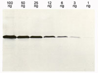
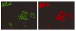
Anti-Histidine-Tagged Protein, mouse monoclonal, clone 13/45/31/2, specifically recognizes an epitage of 6 consecutive His residues. It is validated for use in ELISA, WB, IF & IP.
More>> Anti-Histidine-Tagged Protein, mouse monoclonal, clone 13/45/31/2, specifically recognizes an epitage of 6 consecutive His residues. It is validated for use in ELISA, WB, IF & IP. Less<<MSDS (material safety data sheet) or SDS, CoA and CoQ, dossiers, brochures and other available documents.
Synonyms: Anti-His6
Recommended Products
Overview
| Replacement Information |
|---|
Key Spec Table
| Species Reactivity | Host | Antibody Type |
|---|---|---|
| A | M | Monoclonal Antibody |
Pricing & Availability
| Catalogue Number | Availability | Packaging | Qty/Pack | Price | Quantity | |
|---|---|---|---|---|---|---|
| OB05-100UG |
|
Plastic ampoule | 100 μg |
|
— |
| Product Information | |
|---|---|
| Declaration | Manufactured by Dianova, GmbH, Germany. |
| Form | Liquid |
| Formulation | In 50 mM sodium phosphate buffer, 0.2% gelatin, pH 7.5. |
| Positive control | Proteins containing a His•Tag® sequence |
| Preservative | ≤0.1% sodium azide |
| Quality Level | MQ100 |
| Physicochemical Information |
|---|
| Dimensions |
|---|
| Materials Information |
|---|
| Toxicological Information |
|---|
| Safety Information according to GHS |
|---|
| Safety Information |
|---|
| Product Usage Statements |
|---|
| Storage and Shipping Information | |
|---|---|
| Ship Code | Blue Ice Only |
| Toxicity | Standard Handling |
| Storage | +2°C to +8°C |
| Do not freeze | Yes |
| Packaging Information |
|---|
| Transport Information |
|---|
| Supplemental Information |
|---|
| Specifications |
|---|
| Global Trade Item Number | |
|---|---|
| Catalogue Number | GTIN |
| OB05-100UG | 04055977210057 |
Documentation
Anti-Histidine-Tagged Protein Mouse mAb (13/45/31/2) SDS
| Title |
|---|
Anti-Histidine-Tagged Protein Mouse mAb (13/45/31/2) Certificates of Analysis
| Title | Lot Number |
|---|---|
| OB05 |
References
| Reference overview |
|---|
| Zentgraf, H., et al. 1995. Nucl. Acids. Res. 23, 3347. Gu, J., et al. 1994. BioTechniques 17, 257. Sporeno, E., et al. 1994. J. Biol. Chem., 10991. Sisk, W.P., et al. 1994. J. Virol. 68, 766. Lu, T., et al. 1993. Anal. Biochem. 213, 318. Garner, J., et al. 1992. Cell 69, 833. Hochuli, E., et al. 1987. J. Chromatogr. 411, 177. |