Wenn Sie das Fenster schließen, wird Ihre Konfiguration nicht gespeichert, es sei denn, Sie haben Ihren Artikel in die Bestellung aufgenommen oder zu Ihren Favoriten hinzugefügt.
Klicken Sie auf OK, um das MILLIPLEX® MAP-Tool zu schließen oder auf Abbrechen, um zu Ihrer Auswahl zurückzukehren.
Wählen Sie konfigurierbare Panels & Premixed-Kits - ODER - Kits für die zelluläre Signaltransduktion & MAPmates™
Konfigurieren Sie Ihre MILLIPLEX® MAP-Kits und lassen sich den Preis anzeigen.
Konfigurierbare Panels & Premixed-Kits
Unser breites Angebot enthält Multiplex-Panels, für die Sie die Analyten auswählen können, die am besten für Ihre Anwendung geeignet sind. Unter einem separaten Register können Sie das Premixed-Cytokin-Format oder ein Singleplex-Kit wählen.
Kits für die zelluläre Signaltransduktion & MAPmates™
Wählen Sie gebrauchsfertige Kits zur Erforschung gesamter Signalwege oder Prozesse. Oder konfigurieren Sie Ihre eigenen Kits mit Singleplex MAPmates™.
Die folgenden MAPmates™ sollten nicht zusammen analysiert werden: -MAPmates™, die einen unterschiedlichen Assaypuffer erfordern. -Phosphospezifische und MAPmate™ Gesamtkombinationen wie Gesamt-GSK3β und Gesamt-GSK3β (Ser 9). -PanTyr und locusspezifische MAPmates™, z.B. Phospho-EGF-Rezeptor und Phospho-STAT1 (Tyr701). -Mehr als 1 Phospho-MAPmate™ für ein einziges Target (Akt, STAT3). -GAPDH und β-Tubulin können nicht mit Kits oder MAPmates™, die panTyr enthalten, analysiert werden.
.
Bestellnummer
Bestellinformationen
St./Pkg.
Liste
Dieser Artikel wurde zu Ihren Favoriten hinzugefügt.
Wählen Sie bitte Spezies, Panelart, Kit oder Probenart
Um Ihr MILLIPLEX® MAP-Kit zu konfigurieren, wählen Sie zunächst eine Spezies, eine Panelart und/oder ein Kit.
Custom Premix Selecting "Custom Premix" option means that all of the beads you have chosen will be premixed in manufacturing before the kit is sent to you.
Catalogue Number
Ordering Description
Qty/Pack
List
Dieser Artikel wurde zu Ihren Favoriten hinzugefügt.
Spezies
Panelart
Gewähltes Kit
Menge
Bestellnummer
Bestellinformationen
St./Pkg.
Listenpreis
96-Well Plate
Menge
Bestellnummer
Bestellinformationen
St./Pkg.
Listenpreis
Weitere Reagenzien hinzufügen (MAPmates erfordern die Verwendung eines Puffer- und Detektionskits)
Menge
Bestellnummer
Bestellinformationen
St./Pkg.
Listenpreis
48-602MAG
Buffer Detection Kit for Magnetic Beads
1 Kit
Platzsparende Option Kunden, die mehrere Kits kaufen, können ihre Multiplex-Assaykomponenten in Kunststoffbeuteln anstelle von Packungen erhalten, um eine kompaktere Lagerung zu ermöglichen.
Dieser Artikel wurde zu Ihren Favoriten hinzugefügt.
Das Produkt wurde in Ihre Bestellung aufgenommen
Sie können nun ein weiteres Kit konfigurieren, ein Premixed-Kit wählen, zur Kasse gehen oder das Bestell-Tool schließen.
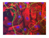
Neurofilaments are a type of intermediate filament that serve as major elements of the cytoskeleton supporting the axon cytoplasm. They are the most abundant fibrillar components of the axon, being on average 3-10 times more frequent than axonal microtubules. Neurofilaments (10nm in dia.) are built from three intertwined protofibrils which are themselves composed of two tetrameric protofilament complexs of monomeric proteins. The neurofilament triplet proteins (68/70, 160, and 200 kDa) occur in both the central and peripheral nervous system and are usually neuron specific. The 68/70 kDa NF-L protein can self-assemble into a filamentous structure, however the 160 kDa NF-M and 200 kDa NF-H proteins require the presence of the 68/70 kDa NF-L protein to co-assemble. Neuromas, ganglioneuromas, gangliogliomas, ganglioneuroblastomas and neuroblastomas stain positively for neurofilaments. Although typically restricted to neurons, neurofilaments have been detected in paragangliomas and adrenal and extra-adrenal pheochromocytomas. Carcinoids, neuroendocrine carcinomas of the skin, and oat cell carcinomas of the lung also express neurofilaments.
Investigator Mini-packs contain smaller qauntities of related antibodies within a specific research area of Neuroscience. The investigator Mini-pack for Neurofilament Antibodies includes monoclonal and polyclonal antibodies against the heavy medium and light chains.
Background Information
Pathway Investigator Antibody MiniPack: Each Pathway Explorer Antibody Minipack contains three related antibodies as part of a signaling cascade or a combination of total and phosphorylated forms of key signaling targets. Each of the three antibodies are 30% the original pack size. Full size versions of each of the Pathway Explorer antibodies are available for sale individually under the same catalog number with the removal of “SP” off of each one (e.g. 05-591SP can be ordered as 05-591).
Neurofilament 70 kDa, clone DA2: Neurofilaments are a type of intermediate filament that serve as major elements of the cytoskeleton supporting the axon cytoplasm. They are the most abundant fibrillar components of the axon, being on average 3-10 times more frequent than axonal microtubules. Neurofilaments (10nm in dia.) are built from three intertwined protofibrils which are themselves composed of two tetrameric protofilament complexs of monomeric proteins. The neurofilament triplet proteins (68/70, 160, and 200 kDa) occur in both the central and peripheral nervous system and are usually neuron specific. The 68/70 kDa NF-L protein can self-assemble into a filamentous structure, however the 160 kDa NF-M and 200 kDa NF-H proteins require the presence of the 68/70 kDa NF-L protein to co-assemble. Neuromas, ganglioneuromas, gangliogliomas, ganglioneuroblastomas and neuroblastomas stain positively for neurofilaments. Although typically restricted to neurons, neurofilaments have been detected in paragangliomas and adrenal and extra-adrenal pheochromocytomas. Carcinoids, neuroendocrine carcinomas of the skin, and oat cell carcinomas of the lung also express neurofilaments.
Neurofilament, Heavy: Neurofilaments are a type of intermediate filament that serve as major elements of the cytoskeleton supporting the axon cytoplasm. They are the most abundant fibrillar components of the axon, being on average 3-10 times more frequent than axonal microtubules. Neurofilaments (10nm in dia.) are built from three intertwined protofibrils which are themselves composed of two tetrameric protofilament complexs of monomeric proteins. The neurofilament triplet proteins (68/70, 160, and 200 kDa) occur in both the central and peripheral nervous system and are usually neuron specific. The 68/70 kDa NF-L protein can self-assemble into a filamentous structure, however the 160 kDa NF-M and 200 kDa NF-H proteins require the presence of the 68/70 kDa NF-L protein to co-assemble. Neuromas, ganglioneuromas, gangliogliomas, ganglioneuroblastomas and neuroblastomas stain positively for neurofilaments. Although typically restricted to neurons, neurofilaments have been detected in paragangliomas and adrenal and extra-adrenal pheochromocytomas. Carcinoids, neuroendocrine carcinomas of the skin, and oat cell carcinomas of the lung also express neurofilaments. * See full size versions for corresponding references.
References
Product Information
Components
MAB1592-C-25UG Anti-Neurofilament NF-Hphos cINP1
AB5539SP Anti-Neurofilament H; 30 μL
MAB1615SP Anti-Neurofilament L; 90μL
Presentation
3 individual tubes containing either Anti-Neurofilament, Heavy; Anti-Neurofilament H & M, phosphorylated, clone NP1; or Anti-Neurofilament 70 kDa, clone DA2
Properties
Each vial is 30% the size of the parent catalog number
FUNCTION: Neurofilaments usually contain three intermediate filament proteins: L, M, and H which are involved in the maintenance of neuronal caliber. NF-H has an important function in mature axons that is not subserved by the two smaller NF proteins. PTM: There are a number of repeats of the tripeptide K-S-P, NFH is phosphorylated on a number of the serines in this motif. It is thought that phosphorylation of NFH results in the formation of interfilament cross bridges that are important in the maintenance of axonal caliber. PTM: Phosphorylation seems to play a major role in the functioning of the larger neurofilament polypeptides (NF-M and NF-H), the levels of phosphorylation being altered developmentally and coincident with a change in the neurofilament function. DISEASE: Defects in NEFH are a cause of susceptibility to amyotrophic lateral sclerosis (ALS) [MIM:105400]. ALS is a neurodegenerative disorder affecting upper and lower motor neurons, and resulting in fatal paralysis. Sensory abnormalities are absent. Death usually occurs within 2 to 5 years. The etiology is likely to be multifactorial, involving both genetic and environmental factors. SIMILARITY: Belongs to the intermediate filament family.
Physicochemical Information
Dimensions
Materials Information
Toxicological Information
Safety Information according to GHS
Safety Information
Product Usage Statements
Quality Assurance
Individual targets are routinely evaluated by Immunohistochemistry and western blot.
Usage Statement
Unless otherwise stated in our catalog or other company documentation accompanying the product(s), our products are intended for research use only and are not to be used for any other purpose, which includes but is not limited to, unauthorized commercial uses, in vitro diagnostic uses, ex vivo or in vivo therapeutic uses or any type of consumption or application to humans or animals.