Wenn Sie das Fenster schließen, wird Ihre Konfiguration nicht gespeichert, es sei denn, Sie haben Ihren Artikel in die Bestellung aufgenommen oder zu Ihren Favoriten hinzugefügt.
Klicken Sie auf OK, um das MILLIPLEX® MAP-Tool zu schließen oder auf Abbrechen, um zu Ihrer Auswahl zurückzukehren.
Wählen Sie konfigurierbare Panels & Premixed-Kits - ODER - Kits für die zelluläre Signaltransduktion & MAPmates™
Konfigurieren Sie Ihre MILLIPLEX® MAP-Kits und lassen sich den Preis anzeigen.
Konfigurierbare Panels & Premixed-Kits
Unser breites Angebot enthält Multiplex-Panels, für die Sie die Analyten auswählen können, die am besten für Ihre Anwendung geeignet sind. Unter einem separaten Register können Sie das Premixed-Cytokin-Format oder ein Singleplex-Kit wählen.
Kits für die zelluläre Signaltransduktion & MAPmates™
Wählen Sie gebrauchsfertige Kits zur Erforschung gesamter Signalwege oder Prozesse. Oder konfigurieren Sie Ihre eigenen Kits mit Singleplex MAPmates™.
Die folgenden MAPmates™ sollten nicht zusammen analysiert werden: -MAPmates™, die einen unterschiedlichen Assaypuffer erfordern. -Phosphospezifische und MAPmate™ Gesamtkombinationen wie Gesamt-GSK3β und Gesamt-GSK3β (Ser 9). -PanTyr und locusspezifische MAPmates™, z.B. Phospho-EGF-Rezeptor und Phospho-STAT1 (Tyr701). -Mehr als 1 Phospho-MAPmate™ für ein einziges Target (Akt, STAT3). -GAPDH und β-Tubulin können nicht mit Kits oder MAPmates™, die panTyr enthalten, analysiert werden.
.
Bestellnummer
Bestellinformationen
St./Pkg.
Liste
Dieser Artikel wurde zu Ihren Favoriten hinzugefügt.
Wählen Sie bitte Spezies, Panelart, Kit oder Probenart
Um Ihr MILLIPLEX® MAP-Kit zu konfigurieren, wählen Sie zunächst eine Spezies, eine Panelart und/oder ein Kit.
Custom Premix Selecting "Custom Premix" option means that all of the beads you have chosen will be premixed in manufacturing before the kit is sent to you.
Catalogue Number
Ordering Description
Qty/Pack
List
Dieser Artikel wurde zu Ihren Favoriten hinzugefügt.
Spezies
Panelart
Gewähltes Kit
Menge
Bestellnummer
Bestellinformationen
St./Pkg.
Listenpreis
96-Well Plate
Menge
Bestellnummer
Bestellinformationen
St./Pkg.
Listenpreis
Weitere Reagenzien hinzufügen (MAPmates erfordern die Verwendung eines Puffer- und Detektionskits)
Menge
Bestellnummer
Bestellinformationen
St./Pkg.
Listenpreis
48-602MAG
Buffer Detection Kit for Magnetic Beads
1 Kit
Platzsparende Option Kunden, die mehrere Kits kaufen, können ihre Multiplex-Assaykomponenten in Kunststoffbeuteln anstelle von Packungen erhalten, um eine kompaktere Lagerung zu ermöglichen.
Dieser Artikel wurde zu Ihren Favoriten hinzugefügt.
Das Produkt wurde in Ihre Bestellung aufgenommen
Sie können nun ein weiteres Kit konfigurieren, ein Premixed-Kit wählen, zur Kasse gehen oder das Bestell-Tool schließen.
ABS1593
Sigma-AldrichAnti-sFRP-1 Antibody
Detect sFRP-1 using this rabbit polyclonal Anti-sFRP-1, Cat. No. ABS1593, validated for use in Immunohistochemistry, Neutralization, and Western Blotting.
More>>Detect sFRP-1 using this rabbit polyclonal Anti-sFRP-1, Cat. No. ABS1593, validated for use in Immunohistochemistry, Neutralization, and Western Blotting. Less<<
Anti-sFRP-1 Antibody: SDB (Sicherheitsdatenblätter), Analysenzertifikate und Qualitätszertifikate, Dossiers, Broschüren und andere verfügbare Dokumente.
Secreted frizzled-related protein 1 (UniProt Q8N474; also known as FRP-1, SARP-2, Secreted apoptosis-related protein 2, sFRP-1) is encoded by the SFRP1 (also known as FRP, FRP1, SARP2) gene (Gene ID 6422) in human. Canonical Wnt signaling is constitutively suppressed in the absence of Wnt, and its activation in the presence of Wnt can be blocked by secreted inhibitors, including the dickkopf-related proteins (DKK) and the secreted frizzled-related proteins (sFRP1-5). sFRPs share homology with the FZD receptor and compete againt FZD for Wnt binding in the extracellular space, sFRPs may also exert their effects by interacting with each other and FZD receptors. sFRP-1 is present during both cardiac development and adult life and is abundantly expressed in the mouse and human heart. Study conducted with Sfrp1-knockout mice indicates a role of sFRP-1 in age-related cardiac deterioration and fibrosis. Human sFRP-1 is produced with a signal peptide sequence (a.a. 1-31), the removal of which yields the mature protein (a.a. 32-314) with an N-terminal FZ (a.a. 53-169) domain, followed by a netrin (NTR) domain (a.a. 186-306).
References
Product Information
Format
Purified
Presentation
Purified rabbit polyclonal antibody in PBS without preservatives.
Detect sFRP-1 using this rabbit polyclonal Anti-sFRP-1, Cat. No. ABS1593, validated for use in Immunohistochemistry, Neutralization, and Western Blotting.
Key Applications
Western Blotting
Immunohistochemistry
Neutralization Assay
Application Notes
Immunohistochemistry Analysis: A 1:260 dilution from a representative lot detected sFRP-1 in human kideny and testis tissue sections (Courtesy of Jeffrey Rubin, M.D., Ph.D., and Aykut Üren, M.D., Ph.D.).
Neutralizing Analysis: A representative lot, in combination with recombinant Wtn5a and an anti-DKK1 antibody, increased migration and mammosphere formation of EpCAM-positive (EPC+) human mammary epithelial cells (MECs) (Scheel, C., et al. (2011). Cell. 145(6):926 940).
Neutralizing Analysis: A representative lot enhanced the formation of TRACP+ mutinucleated and mononucleated cells from co-cultured murine splenic and primary osteoblastic cells upon PGE2 and 1 ,25 (OH)2D3 stimulatation (Häusler, K.D., et al. (2004). J. Bone Miner. Res. 19(11):1873-1881).
Note: Detection of endogenous sFRP-1 in whole cell lysates or tissue samples is difficult without an enrichment step prior to immunoblotting (e.g. by immunoprecipitation or heparin affinity chromatography).
Biological Information
Immunogen
Mammalian expressed human sFRP-1, purified from condition culture medium (Häusler, K.D., et al. (2004). J. Bone Miner. Res. 19(11):1873-1881; Uren, A., et al. (2000). J. Biol. Chem. 275(6):4374-4382).
Concentration
Please refer to lot specific datasheet.
Host
Rabbit
Specificity
This polyclonal antibody neutralized the inhibitory activity of sFRP-1 (Scheel, C., et al. (2011). Cell. 145(6):926 940; Häusler, K.D., et al. (2004). J. Bone Miner. Res. 19(11):1873-1881).
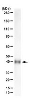
~39 kDa observed. Target band size appears larger than the calculated molecular weights of 32.54/35.39 kDa (mature/pro-form) due to glycosylation. Uncharacterized bands may be observed in some lysate(s).
Physicochemical Information
Dimensions
Materials Information
Toxicological Information
Safety Information according to GHS
Safety Information
Product Usage Statements
Quality Assurance
Evaluated by Western Blotting of human sFRP-1 recombinant protein.
Western Blotting Analysis: 1 µg/mL of this antibody detected in 10 ng of mammalian expressed, secreted human sFRP-1.
Usage Statement
Unless otherwise stated in our catalog or other company documentation accompanying the product(s), our products are intended for research use only and are not to be used for any other purpose, which includes but is not limited to, unauthorized commercial uses, in vitro diagnostic uses, ex vivo or in vivo therapeutic uses or any type of consumption or application to humans or animals.
Storage and Shipping Information
Storage Conditions
Stable for 1 year at -20°C from date of receipt. Handling Recommendations: Upon receipt and prior to removing the cap, centrifuge the vial and gently mix the solution. Aliquot into microcentrifuge tubes and store at -20°C. Avoid repeated freeze/thaw cycles, which may damage IgG and affect product performance.