Wenn Sie das Fenster schließen, wird Ihre Konfiguration nicht gespeichert, es sei denn, Sie haben Ihren Artikel in die Bestellung aufgenommen oder zu Ihren Favoriten hinzugefügt.
Klicken Sie auf OK, um das MILLIPLEX® MAP-Tool zu schließen oder auf Abbrechen, um zu Ihrer Auswahl zurückzukehren.
Wählen Sie konfigurierbare Panels & Premixed-Kits - ODER - Kits für die zelluläre Signaltransduktion & MAPmates™
Konfigurieren Sie Ihre MILLIPLEX® MAP-Kits und lassen sich den Preis anzeigen.
Konfigurierbare Panels & Premixed-Kits
Unser breites Angebot enthält Multiplex-Panels, für die Sie die Analyten auswählen können, die am besten für Ihre Anwendung geeignet sind. Unter einem separaten Register können Sie das Premixed-Cytokin-Format oder ein Singleplex-Kit wählen.
Kits für die zelluläre Signaltransduktion & MAPmates™
Wählen Sie gebrauchsfertige Kits zur Erforschung gesamter Signalwege oder Prozesse. Oder konfigurieren Sie Ihre eigenen Kits mit Singleplex MAPmates™.
Die folgenden MAPmates™ sollten nicht zusammen analysiert werden: -MAPmates™, die einen unterschiedlichen Assaypuffer erfordern. -Phosphospezifische und MAPmate™ Gesamtkombinationen wie Gesamt-GSK3β und Gesamt-GSK3β (Ser 9). -PanTyr und locusspezifische MAPmates™, z.B. Phospho-EGF-Rezeptor und Phospho-STAT1 (Tyr701). -Mehr als 1 Phospho-MAPmate™ für ein einziges Target (Akt, STAT3). -GAPDH und β-Tubulin können nicht mit Kits oder MAPmates™, die panTyr enthalten, analysiert werden.
.
Bestellnummer
Bestellinformationen
St./Pkg.
Liste
Dieser Artikel wurde zu Ihren Favoriten hinzugefügt.
Wählen Sie bitte Spezies, Panelart, Kit oder Probenart
Um Ihr MILLIPLEX® MAP-Kit zu konfigurieren, wählen Sie zunächst eine Spezies, eine Panelart und/oder ein Kit.
Custom Premix Selecting "Custom Premix" option means that all of the beads you have chosen will be premixed in manufacturing before the kit is sent to you.
Catalogue Number
Ordering Description
Qty/Pack
List
Dieser Artikel wurde zu Ihren Favoriten hinzugefügt.
Spezies
Panelart
Gewähltes Kit
Menge
Bestellnummer
Bestellinformationen
St./Pkg.
Listenpreis
96-Well Plate
Menge
Bestellnummer
Bestellinformationen
St./Pkg.
Listenpreis
Weitere Reagenzien hinzufügen (MAPmates erfordern die Verwendung eines Puffer- und Detektionskits)
Menge
Bestellnummer
Bestellinformationen
St./Pkg.
Listenpreis
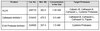
48-602MAG
Buffer Detection Kit for Magnetic Beads
1 Kit
Platzsparende Option Kunden, die mehrere Kits kaufen, können ihre Multiplex-Assaykomponenten in Kunststoffbeuteln anstelle von Packungen erhalten, um eine kompaktere Lagerung zu ermöglichen.
Dieser Artikel wurde zu Ihren Favoriten hinzugefügt.
Das Produkt wurde in Ihre Bestellung aufgenommen
Sie können nun ein weiteres Kit konfigurieren, ein Premixed-Kit wählen, zur Kasse gehen oder das Bestell-Tool schließen.
Xeroderma pigmentosum group B-complementing protein
XPB
Background Information
Transcription factor II human (TFIIH) basal transcription factor complex helicase XPB subunit (EC 3.6.4.12; UniProt P19447; also known as BTF2 p89, DNA excision repair protein ERCC-3, DNA repair helicase, DNA repair protein complementing XP-B cells, TFIIH 89 kDa subunit, TFIIH basal transcription factor complex 89 kDa subunit, TFIIH p89, Basic transcription factor 2 89 kDa subunit, Xeroderma pigmentosum group B-complementing protein) is encoded by the ERCC3 (also known as BTF2, GTF2H, RAD25, TFIIH, XPB) gene (Gene ID 2071) in human. DNA lesions caused by UV irradiation, drugs, or other environmental factors are eliminated by two nucleotide excision repair (NER) pathways, Global genome repair (GGR) and transcription-coupled repair (TCR). In GGR, the removal of lesions requires their recognition by the repair factor XPC/HR23b and the subsequent opening of the DNA duplex by TFIIH. The resulting single-stranded structure is stabilized by XPA and replication protein A (RPA). XPG is recruited through its interaction with TFIIH on the 3′ side of the lesion and its positioning on the cut site requires RPA. The interaction between XPA and XPB (ERCC1) stimulates the recruitment of ERCC1-XPF on the 5′ side of the DNA lesion. The damaged oligonucleotide can then be removed through the double incision by XPG and ERCC1-XPF endonucleases. In TCR, these factors (except XPC/HR23B) are recruited by the stalled RNA pol II in front of the damage with the help of the CSB and CSA proteins.
References
Product Information
Format
Ascites
Presentation
Mouse monoclonal IgG1κ ascites with 0.05% sodium azide.
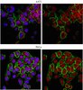
This Anti-XPB Antibody, clone 15TF2-1B3 is validated for use in Western Blotting, Immunocytochemistry for the detection of XPB.
Key Applications
Western Blotting
Immunocytochemistry
Application Notes
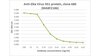
Western Blotting Analysis: A representative lot detected Xpb in murine embryonic fibroblasts (MEFs) and HeLa cells, as well as in transgenic animal-derived MEFs expressing Xpb lacking last 43 C-terminal amino acids (Andressoo, J.O., et al. (2009). Mol Cell Biol.29(5):1276-290). Western Blotting Analysis: A representative lot detected endougenous as well as exogenously expressed Xpb in both U2OS17 whole cell lysate and in THIIF p62 subunit immunoprecipitate (Ziani, S., et al. (2014). J Cell Biol.;206(5):589-598). Western Blotting Analysis: A representative lot detected Xpb in THIIF TTDA subunit immunoprecipitate (Giglia-Mari,G., et al. (2006). PLoS Biol. 4(6): e156). Immunocytochemistry Analysis: A representative lot detected XPB recruitment to the DNA damage sites in the nuclei of UV-irradated HeLa cells (Alekseev, S., et al. (2014). Chem Biol. 21(3):398-407).
~85 kDa observed. Uncharacterized band(s) may appear in some lysates.
Physicochemical Information
Dimensions
Materials Information
Toxicological Information
Safety Information according to GHS
Safety Information
Product Usage Statements
Quality Assurance
Evaluated by Western Blotting in HeLa nuclear extract.
Western Blotting Analysis: A 1:2,000 dilution of this antibody detected XPB in 10 µg of HeLa nuclear extract.
Usage Statement
Unless otherwise stated in our catalog or other company documentation accompanying the product(s), our products are intended for research use only and are not to be used for any other purpose, which includes but is not limited to, unauthorized commercial uses, in vitro diagnostic uses, ex vivo or in vivo therapeutic uses or any type of consumption or application to humans or animals.
Storage and Shipping Information
Storage Conditions
Stable for 1 year at -20°C from date of receipt. Handling Recommendations: Upon receipt and prior to removing the cap, centrifuge the vial and gently mix the solution. Aliquot into microcentrifuge tubes and store at -20°C. Avoid repeated freeze/thaw cycles, which may damage IgG and affect product performance.
Sequential and ordered assembly of a large DNA repair complex on undamaged chromatin. Ziani, S; Nagy, Z; Alekseev, S; Soutoglou, E; Egly, JM; Coin, F The Journal of cell biology
206
589-98
2014
In nucleotide excision repair (NER), damage recognition by XPC-hHR23b is described as a critical step in the formation of the preincision complex (PInC) further composed of TFIIH, XPA, RPA, XPG, and ERCC1-XPF. To obtain new molecular insights into the assembly of the PInC, we analyzed its formation independently of DNA damage by using the lactose operator/repressor reporter system. We observed a sequential and ordered self-assembly of the PInC operating upon immobilization of individual NER factors on undamaged chromatin and mimicking that functioning on a bona fide NER substrate. We also revealed that the recruitment of the TFIIH subunit TTDA, involved in trichothiodystrophy group A disorder (TTD-A), was key in the completion of the PInC. TTDA recruits XPA through its first 15 amino acids, depleted in some TTD-A patients. More generally, these results show that proteins forming large nuclear complexes can be recruited sequentially on chromatin in the absence of their natural DNA target and with no reciprocity in their recruitment.
A small molecule screen identifies an inhibitor of DNA repair inducing the degradation of TFIIH and the chemosensitization of tumor cells to platinum. Alekseev, S; Ayadi, M; Brino, L; Egly, JM; Larsen, AK; Coin, F Chemistry & biology
21
398-407
2014
Nucleotide excision repair (NER) removes DNA lesions resulting from exposure to UV irradiation or chemical agents such as platinum-based drugs used as anticancer molecules. Pharmacological inhibition of NER is expected to enhance chemosensitivity but nontoxic NER inhibitors are rare. Using a drug repositioning approach, we identify spironolactone (SP), an antagonist of aldosterone, as a potent NER inhibitor. We found that SP promotes a rapid and reversible degradation of XPB, a subunit of transcription/repair factor TFIIH. Such degradation depends both on ubiquitin-activating enzyme and on the 26S proteasome. Supplementation of extracts from SP-treated cells with purified TFIIH restored TFIIH-dependent repair and transcription activities in vitro, demonstrating the specific impact of SP on two fundamental functions of TFIIH. Finally, SP potentiated the cytotoxicity of platinum derivatives toward tumor cells, making it a potential therapeutic and research tool.
An Xpb mouse model for combined xeroderma pigmentosum and cockayne syndrome reveals progeroid features upon further attenuation of DNA repair. Andressoo, JO; Weeda, G; de Wit, J; Mitchell, JR; Beems, RB; van Steeg, H; van der Horst, GT; Hoeijmakers, JH Molecular and cellular biology
29
1276-90
2009
Patients carrying mutations in the XPB helicase subunit of the basal transcription and nucleotide excision repair (NER) factor TFIIH display the combined cancer and developmental-progeroid disorder xeroderma pigmentosum/Cockayne syndrome (XPCS). Due to the dual transcription repair role of XPB and the absence of animal models, the underlying molecular mechanisms of XPB(XPCS) are largely uncharacterized. Here we show that severe alterations in Xpb cause embryonic lethality and that knock-in mice closely mimicking an XPCS patient-derived XPB mutation recapitulate the UV sensitivity typical for XP but fail to show overt CS features unless the DNA repair capacity is further challenged by crossings to the NER-deficient Xpa background. Interestingly, the Xpb(XPCS) Xpa double mutants display a remarkable interanimal variance, which points to stochastic DNA damage accumulation as an important determinant of clinical diversity in NER syndromes. Furthermore, mice carrying the Xpb(XPCS) mutation together with a point mutation in the second TFIIH helicase Xpd are healthy at birth but display neonatal lethality, indicating that transcription efficiency is sufficient to permit embryonal development even when both TFIIH helicases are crippled. The double-mutant cells exhibit sensitivity to oxidative stress, suggesting a role for endogenous DNA damage in the onset of XPB-associated CS.
Transcription/repair factor IIH (TFIIH) is essential for RNA polymerase II transcription and nucleotide excision repair (NER). This multi-subunit complex consists of ten polypeptides, including the recently identified small 8-kDa trichothiodystrophy group A (TTDA)/ hTFB5 protein. Patients belonging to the rare neurodevelopmental repair syndrome TTD-A carry inactivating mutations in the TTDA/hTFB5 gene. One of these mutations completely inactivates the protein, whereas other TFIIH genes only tolerate point mutations that do not compromise the essential role in transcription. Nevertheless, the severe NER-deficiency in TTD-A suggests that the TTDA protein is critical for repair. Using a fluorescently tagged and biologically active version of TTDA, we have investigated the involvement of TTDA in repair and transcription in living cells. Under non-challenging conditions, TTDA is present in two distinct kinetic pools: one bound to TFIIH, and a free fraction that shuttles between the cytoplasm and nucleus. After induction of NER-specific DNA lesions, the equilibrium between these two pools dramatically shifts towards a more stable association of TTDA to TFIIH. Modulating transcriptional activity in cells did not induce a similar shift in this equilibrium. Surprisingly, DNA conformations that only provoke an abortive-type of NER reaction do not result into a more stable incorporation of TTDA into TFIIH. These findings identify TTDA as the first TFIIH subunit with a primarily NER-dedicated role in vivo and indicate that its interaction with TFIIH reflects productive NER.