Wenn Sie das Fenster schließen, wird Ihre Konfiguration nicht gespeichert, es sei denn, Sie haben Ihren Artikel in die Bestellung aufgenommen oder zu Ihren Favoriten hinzugefügt.
Klicken Sie auf OK, um das MILLIPLEX® MAP-Tool zu schließen oder auf Abbrechen, um zu Ihrer Auswahl zurückzukehren.
Wählen Sie konfigurierbare Panels & Premixed-Kits - ODER - Kits für die zelluläre Signaltransduktion & MAPmates™
Konfigurieren Sie Ihre MILLIPLEX® MAP-Kits und lassen sich den Preis anzeigen.
Konfigurierbare Panels & Premixed-Kits
Unser breites Angebot enthält Multiplex-Panels, für die Sie die Analyten auswählen können, die am besten für Ihre Anwendung geeignet sind. Unter einem separaten Register können Sie das Premixed-Cytokin-Format oder ein Singleplex-Kit wählen.
Kits für die zelluläre Signaltransduktion & MAPmates™
Wählen Sie gebrauchsfertige Kits zur Erforschung gesamter Signalwege oder Prozesse. Oder konfigurieren Sie Ihre eigenen Kits mit Singleplex MAPmates™.
Die folgenden MAPmates™ sollten nicht zusammen analysiert werden: -MAPmates™, die einen unterschiedlichen Assaypuffer erfordern. -Phosphospezifische und MAPmate™ Gesamtkombinationen wie Gesamt-GSK3β und Gesamt-GSK3β (Ser 9). -PanTyr und locusspezifische MAPmates™, z.B. Phospho-EGF-Rezeptor und Phospho-STAT1 (Tyr701). -Mehr als 1 Phospho-MAPmate™ für ein einziges Target (Akt, STAT3). -GAPDH und β-Tubulin können nicht mit Kits oder MAPmates™, die panTyr enthalten, analysiert werden.
.
Bestellnummer
Bestellinformationen
St./Pkg.
Liste
Dieser Artikel wurde zu Ihren Favoriten hinzugefügt.
Wählen Sie bitte Spezies, Panelart, Kit oder Probenart
Um Ihr MILLIPLEX® MAP-Kit zu konfigurieren, wählen Sie zunächst eine Spezies, eine Panelart und/oder ein Kit.
Custom Premix Selecting "Custom Premix" option means that all of the beads you have chosen will be premixed in manufacturing before the kit is sent to you.
Catalogue Number
Ordering Description
Qty/Pack
List
Dieser Artikel wurde zu Ihren Favoriten hinzugefügt.
Spezies
Panelart
Gewähltes Kit
Menge
Bestellnummer
Bestellinformationen
St./Pkg.
Listenpreis
96-Well Plate
Menge
Bestellnummer
Bestellinformationen
St./Pkg.
Listenpreis
Weitere Reagenzien hinzufügen (MAPmates erfordern die Verwendung eines Puffer- und Detektionskits)
Menge
Bestellnummer
Bestellinformationen
St./Pkg.
Listenpreis
48-602MAG
Buffer Detection Kit for Magnetic Beads
1 Kit
Platzsparende Option Kunden, die mehrere Kits kaufen, können ihre Multiplex-Assaykomponenten in Kunststoffbeuteln anstelle von Packungen erhalten, um eine kompaktere Lagerung zu ermöglichen.
Dieser Artikel wurde zu Ihren Favoriten hinzugefügt.
Das Produkt wurde in Ihre Bestellung aufgenommen
Sie können nun ein weiteres Kit konfigurieren, ein Premixed-Kit wählen, zur Kasse gehen oder das Bestell-Tool schließen.
Anti-THAP11 Antibody, clone 3F3 is a highly specific mouse monoclonal antibody, that targets Thanatos-associated protein 11 & has been tested in western blotting, Immunofluorescence, IHC & Flow Cytometry.
More>>Anti-THAP11 Antibody, clone 3F3 is a highly specific mouse monoclonal antibody, that targets Thanatos-associated protein 11 & has been tested in western blotting, Immunofluorescence, IHC & Flow Cytometry. Less<<
Anti-THAP11 Antibody, clone 3F3: SDB (Sicherheitsdatenblätter), Analysenzertifikate und Qualitätszertifikate, Dossiers, Broschüren und andere verfügbare Dokumente.
THAP11 or Thanatos-associated protein 11, THAP domain-containing protein 11, encoded by the human gene named THAP11 or HRIHFB2206 is a sequence specific transcription factor critical for embryogenesis and the pluripotency of embryonic stem cells (ESCs). THAP11 is a transcriptional repressor that suppresses differentiation genes in ES cells by suppressing c-Myc. Overexpression of THAP11 markedly inhibits growth of a number of different cells, including some cancer cells and non-transformed cells. THAP11 is ubiquitously expressed in normal tissues and frequently downregulated in several human tumor tissues. Research demonstrates that among the cellular protein partners of THAP11 is a transcriptional activator PCBP1, where again overexpression inhibited CD44v6 expression (one of the homing receptors) and cell invasion in the cancer cell line HepG2. However THAP11 is not always associated with growth suppression as recent research in colon cancer indicates that THAP11 associates with the transcriptional coregulator HCF-1 (host cell factor 1) and recruits HCF-1 to target promoters leading to an increase in gene expression. Moreover THAP11 is found to be expressed in both primary tumors and metastases, and knockdown of THAP11 in SW620 colon cancer cells lines showed a marked decrease in cell proliferation and a down regulation of numerous genes by gene profiling. EMD-Millipore’s Anti-THAP11 mouse monoclonal antibody has been tested in western blot against HeLa and NTERA-2 cell lysates and paraffin embedded immunohistochemistry on colon and ovarian cancer tissue sections and in fluorescent immunocytochemistry on NTERA-2 cells in culture and in flow cytometry on HeLa cells in culture.
References
Product Information
Format
Ascites
Control
HeLa and NTERA-2 cell lysates
Presentation
Mouse monoclonal IgG1 ascitic fluid containing up to 0.1% sodium azide.
Anti-THAP11 Antibody, clone 3F3 is a highly specific mouse monoclonal antibody, that targets Thanatos-associated protein 11 & has been tested in western blotting, Immunofluorescence, IHC & Flow Cytometry.
Key Applications
Western Blotting
Immunofluorescence
Immunohistochemistry
Flow Cytometry
Application Notes
Immunofluorescence Analysis: A 1:200-1,000 dilution from a representative lot detected THAP11 in NTERA-2 cells.
Immunohistochemistry Analysis: A 1:200-1,000 dilution from a representative lot detected THAP11 in paraffin-embedded colon cancer and ovarian tissues.
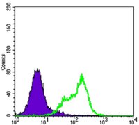
Flow Cytometry Analysis: A 1:200-400 dilution from a representative lot detected THAP11 in HeLa cells.
Optimal working dilutions must be determined by end user.
Biological Information
Immunogen
Purified recombinant fragment of human THAP11 expressed in E. Coli.
~48 kDa observed. Uncharacterized bands may appear in some lysate(s).
Physicochemical Information
Dimensions
Materials Information
Toxicological Information
Safety Information according to GHS
Safety Information
Product Usage Statements
Quality Assurance
Evaluated by Western Blotting in HeLa and NTERA-2 cell lysates.
Western Blotting Analysis: A 1:500-2,000 dilution of this antibody detected THAP11 in HeLa and NTERA-2 cell lysates.
Usage Statement
Unless otherwise stated in our catalog or other company documentation accompanying the product(s), our products are intended for research use only and are not to be used for any other purpose, which includes but is not limited to, unauthorized commercial uses, in vitro diagnostic uses, ex vivo or in vivo therapeutic uses or any type of consumption or application to humans or animals.
Storage and Shipping Information
Storage Conditions
Stable for 1 year at -20°C from date of receipt. Handling Recommendations: Upon receipt and prior to removing the cap, centrifuge the vial and gently mix the solution. Aliquot into microcentrifuge tubes and store at -20°C. Avoid repeated freeze/thaw cycles, which may damage IgG and affect product performance.