Wenn Sie das Fenster schließen, wird Ihre Konfiguration nicht gespeichert, es sei denn, Sie haben Ihren Artikel in die Bestellung aufgenommen oder zu Ihren Favoriten hinzugefügt.
Klicken Sie auf OK, um das MILLIPLEX® MAP-Tool zu schließen oder auf Abbrechen, um zu Ihrer Auswahl zurückzukehren.
Wählen Sie konfigurierbare Panels & Premixed-Kits - ODER - Kits für die zelluläre Signaltransduktion & MAPmates™
Konfigurieren Sie Ihre MILLIPLEX® MAP-Kits und lassen sich den Preis anzeigen.
Konfigurierbare Panels & Premixed-Kits
Unser breites Angebot enthält Multiplex-Panels, für die Sie die Analyten auswählen können, die am besten für Ihre Anwendung geeignet sind. Unter einem separaten Register können Sie das Premixed-Cytokin-Format oder ein Singleplex-Kit wählen.
Kits für die zelluläre Signaltransduktion & MAPmates™
Wählen Sie gebrauchsfertige Kits zur Erforschung gesamter Signalwege oder Prozesse. Oder konfigurieren Sie Ihre eigenen Kits mit Singleplex MAPmates™.
Die folgenden MAPmates™ sollten nicht zusammen analysiert werden: -MAPmates™, die einen unterschiedlichen Assaypuffer erfordern. -Phosphospezifische und MAPmate™ Gesamtkombinationen wie Gesamt-GSK3β und Gesamt-GSK3β (Ser 9). -PanTyr und locusspezifische MAPmates™, z.B. Phospho-EGF-Rezeptor und Phospho-STAT1 (Tyr701). -Mehr als 1 Phospho-MAPmate™ für ein einziges Target (Akt, STAT3). -GAPDH und β-Tubulin können nicht mit Kits oder MAPmates™, die panTyr enthalten, analysiert werden.
.
Bestellnummer
Bestellinformationen
St./Pkg.
Liste
Dieser Artikel wurde zu Ihren Favoriten hinzugefügt.
Wählen Sie bitte Spezies, Panelart, Kit oder Probenart
Um Ihr MILLIPLEX® MAP-Kit zu konfigurieren, wählen Sie zunächst eine Spezies, eine Panelart und/oder ein Kit.
Custom Premix Selecting "Custom Premix" option means that all of the beads you have chosen will be premixed in manufacturing before the kit is sent to you.
Catalogue Number
Ordering Description
Qty/Pack
List
Dieser Artikel wurde zu Ihren Favoriten hinzugefügt.
Spezies
Panelart
Gewähltes Kit
Menge
Bestellnummer
Bestellinformationen
St./Pkg.
Listenpreis
96-Well Plate
Menge
Bestellnummer
Bestellinformationen
St./Pkg.
Listenpreis
Weitere Reagenzien hinzufügen (MAPmates erfordern die Verwendung eines Puffer- und Detektionskits)
Menge
Bestellnummer
Bestellinformationen
St./Pkg.
Listenpreis
48-602MAG
Buffer Detection Kit for Magnetic Beads
1 Kit
Platzsparende Option Kunden, die mehrere Kits kaufen, können ihre Multiplex-Assaykomponenten in Kunststoffbeuteln anstelle von Packungen erhalten, um eine kompaktere Lagerung zu ermöglichen.
Dieser Artikel wurde zu Ihren Favoriten hinzugefügt.
Das Produkt wurde in Ihre Bestellung aufgenommen
Sie können nun ein weiteres Kit konfigurieren, ein Premixed-Kit wählen, zur Kasse gehen oder das Bestell-Tool schließen.
Anti-REP-1 Antibody, clone 2F1 is an antibody against REP-1 for use in WB, IP, IC, IH.
More>>Anti-REP-1 Antibody, clone 2F1 is an antibody against REP-1 for use in WB, IP, IC, IH. Less<<
Anti-REP-1 Antibody, clone 2F1: SDB (Sicherheitsdatenblätter), Analysenzertifikate und Qualitätszertifikate, Dossiers, Broschüren und andere verfügbare Dokumente.
Rab proteins are low molecular weight, ras-related GTPases that bind to a cell membrane’s cytoplasmic surface and function to regulate protein trafficking in both endocytic and secretory pathways. Rab escort protein (REP)-1 is a 653 amino acid protein belonging to the Rab GDI family of proteins. REP-1 binds to newly assembled unprenylated RAB proteins, escorts them to the Rab GGTases, and remains attached during and after the geranylgeranyl transfer reaction. REP proteins are composed of two conserved domains connected by a 150 amino acid insert. The multisheet domain I acts to assemble a Rab-binding platform. A globular, smaller, α-helical domain II, is thought to participate in membrane-protein interaction. Defects in the expression of REP-1 cause truncation or absence of the protein, and result in a disease called choroideremia (CHM), an X-linked blindness characterized by a progressive dystrophy of the retinal pigment epithelium, retina, and the choroid.
References
Product Information
Format
Purified
Control
293T cell lysate
Presentation
Purified mouse monoclonal IgG1κ in buffer containing 0.1 M Tris-Glycine (pH 7.4), 150 mM NaCl with 0.05% sodium azide.
Anti-REP-1 Antibody, clone 2F1 is an antibody against REP-1 for use in WB, IP, IC, IH.
Key Applications
Western Blotting
Immunoprecipitation
Immunocytochemistry
Immunohistochemistry
Application Notes
Immunohistochemistry Analysis: A previous lot of MABN52 was successfully used in IH, as reported by an independent laboratory (MacDonald, I.M., et al. (2005). Invest Ophthalmol Vis Sci. 46:E-Abstract 540).
Biological Information
Immunogen
Recombinant protein corresponding to human REP-1.
Epitope
Unknown
Clone
2F1
Concentration
Please refer to the Certificate of Analysis for the lot-specific concentration.
This gene encodes component A of the RAB geranylgeranyl transferase holoenzyme. In the dimeric holoenzyme, this subunit binds unprenylated Rab GTPases and then presents them to the catalytic Rab GGTase subunit for the geranylgeranyl transfer reaction. Rab GTPases need to be geranylgeranyled on either one or two cysteine residues in their C-terminus to localize to the correct intracellular membrane. Mutations in this gene are a cause of choroideremia; also known as tapetochoroidal dystrophy (TCD). This X-linked disease is characterized by progressive dystrophy of the choroid, retinal pigment epithelium and retina. Alternative splicing results in multiple transcript variants encoding different isoforms.
FUNCTION: Binds unprenylated Rab proteins, presents it to the catalytic Rab GGTase dimer, and remains bound to it after the geranylgeranyl transfer reaction. The component A is thought to be regenerated by transferring its prenylated Rab back to the donor membrane.
SUBUNIT STRUCTURE: Monomer. Interacts with RAB1B; the interaction is required for RAB1B prenylation. Interacts with RGGT (By similarity).
INVOLVEMENT IN DISEASE: Defects in CHM are the cause of choroideremia (CHM) [MIM:303100]; also known as tapetochoroidal dystrophy (TCD). CHM is a common form of X-linked blindness characterized by progressive dystrophy of the choroid, retinal pigment epithelium and retina.
SEQUENCE SIMLARITIES: Belongs to the Rab GDI family.
Molecular Weight
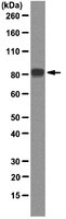
~83 kDa observed
Physicochemical Information
Dimensions
Materials Information
Toxicological Information
Safety Information according to GHS
Safety Information
Product Usage Statements
Quality Assurance
Evaluated by Western Blot in 293T cell lysate.
Western Blot Analysis: 0.5 µg/mL of this antibody detected REP-1 on 10 µg of 293T cell lysate.
Usage Statement
Unless otherwise stated in our catalog or other company documentation accompanying the product(s), our products are intended for research use only and are not to be used for any other purpose, which includes but is not limited to, unauthorized commercial uses, in vitro diagnostic uses, ex vivo or in vivo therapeutic uses or any type of consumption or application to humans or animals.