Wenn Sie das Fenster schließen, wird Ihre Konfiguration nicht gespeichert, es sei denn, Sie haben Ihren Artikel in die Bestellung aufgenommen oder zu Ihren Favoriten hinzugefügt.
Klicken Sie auf OK, um das MILLIPLEX® MAP-Tool zu schließen oder auf Abbrechen, um zu Ihrer Auswahl zurückzukehren.
Wählen Sie konfigurierbare Panels & Premixed-Kits - ODER - Kits für die zelluläre Signaltransduktion & MAPmates™
Konfigurieren Sie Ihre MILLIPLEX® MAP-Kits und lassen sich den Preis anzeigen.
Konfigurierbare Panels & Premixed-Kits
Unser breites Angebot enthält Multiplex-Panels, für die Sie die Analyten auswählen können, die am besten für Ihre Anwendung geeignet sind. Unter einem separaten Register können Sie das Premixed-Cytokin-Format oder ein Singleplex-Kit wählen.
Kits für die zelluläre Signaltransduktion & MAPmates™
Wählen Sie gebrauchsfertige Kits zur Erforschung gesamter Signalwege oder Prozesse. Oder konfigurieren Sie Ihre eigenen Kits mit Singleplex MAPmates™.
Die folgenden MAPmates™ sollten nicht zusammen analysiert werden: -MAPmates™, die einen unterschiedlichen Assaypuffer erfordern. -Phosphospezifische und MAPmate™ Gesamtkombinationen wie Gesamt-GSK3β und Gesamt-GSK3β (Ser 9). -PanTyr und locusspezifische MAPmates™, z.B. Phospho-EGF-Rezeptor und Phospho-STAT1 (Tyr701). -Mehr als 1 Phospho-MAPmate™ für ein einziges Target (Akt, STAT3). -GAPDH und β-Tubulin können nicht mit Kits oder MAPmates™, die panTyr enthalten, analysiert werden.
.
Bestellnummer
Bestellinformationen
St./Pkg.
Liste
Dieser Artikel wurde zu Ihren Favoriten hinzugefügt.
Wählen Sie bitte Spezies, Panelart, Kit oder Probenart
Um Ihr MILLIPLEX® MAP-Kit zu konfigurieren, wählen Sie zunächst eine Spezies, eine Panelart und/oder ein Kit.
Custom Premix Selecting "Custom Premix" option means that all of the beads you have chosen will be premixed in manufacturing before the kit is sent to you.
Catalogue Number
Ordering Description
Qty/Pack
List
Dieser Artikel wurde zu Ihren Favoriten hinzugefügt.
Spezies
Panelart
Gewähltes Kit
Menge
Bestellnummer
Bestellinformationen
St./Pkg.
Listenpreis
96-Well Plate
Menge
Bestellnummer
Bestellinformationen
St./Pkg.
Listenpreis
Weitere Reagenzien hinzufügen (MAPmates erfordern die Verwendung eines Puffer- und Detektionskits)
Menge
Bestellnummer
Bestellinformationen
St./Pkg.
Listenpreis
48-602MAG
Buffer Detection Kit for Magnetic Beads
1 Kit
Platzsparende Option Kunden, die mehrere Kits kaufen, können ihre Multiplex-Assaykomponenten in Kunststoffbeuteln anstelle von Packungen erhalten, um eine kompaktere Lagerung zu ermöglichen.
Dieser Artikel wurde zu Ihren Favoriten hinzugefügt.
Das Produkt wurde in Ihre Bestellung aufgenommen
Sie können nun ein weiteres Kit konfigurieren, ein Premixed-Kit wählen, zur Kasse gehen oder das Bestell-Tool schließen.
Anti-PHF1, clone 12C2.1, Cat. No. MABE676, is a highly specific mouse monoclonal antibody that targets PHF1 and has been tested in Immunohistochemistry (Paraffin) and Western Blotting.
More>>Anti-PHF1, clone 12C2.1, Cat. No. MABE676, is a highly specific mouse monoclonal antibody that targets PHF1 and has been tested in Immunohistochemistry (Paraffin) and Western Blotting. Less<<
Anti-PHF1 Antibody, clone 12C2.1: SDB (Sicherheitsdatenblätter), Analysenzertifikate und Qualitätszertifikate, Dossiers, Broschüren und andere verfügbare Dokumente.
PHD finger protein 1 (UniProt: O43189; also known as PHF1, hPHF1, Polycomb-like protein 1, hPCl1) is encoded by the PHF1 (also known as PCL1) gene (Gene ID: 5252) in human. PHF1 is a member of the polycomblike family and contains two PHD-type zinc fingers (aa 87-142 and 186-240) and one Tudor domain (aa 29-86). It is essential for epigenetic regulation and genome maintenance. It specifically binds histone H3 trimethylated at 'Lys-36' (H3K36me3) via its Tudor domain and recruits the PRC2 complex. PHF1 is highly expressed in heart, skeletal muscle, and pancreas, with lower levels found in brain, placenta, lung, liver and kidney. It is involved in DNA damage response and is recruited at double-strand breaks. PHF1 localizes to the promoters of numerous target genes and to double-strand breaks (DSBs) sites following DNA damage. It has been reported that PRC2 recruitment by PHF1 promotes H3K27me3 and subsequent gene silencing by inducing spreading of PRC2 and H3K27me3 into H3K36me3 loci. A chromosomal aberration involving PHF1 is reported to be the causative factor in endometrial stromal tumors. (Ref.: Sarma, K et al. (2008). Mol. Cell. Biol. 28(8): 2718-31; Musselman, CA et al (2012). Nat. Struct. Mol. Biol. 19(12):1266-72).
References
Product Information
Format
Purified
Presentation
Purified mouse monoclonal antibody IgG2b in buffer containing 0.1 M Tris-Glycine (pH 7.4), 150 mM NaCl with 0.05% sodium azide.
Anti-PHF1, clone 12C2.1, Cat. No. MABE676, is a highly specific mouse monoclonal antibody that targets PHF1 and has been tested in Immunohistochemistry (Paraffin) and Western Blotting.
Key Applications
Western Blotting
Immunohistochemistry (Paraffin)
Application Notes
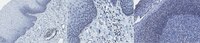
Immunohistochemistry Analysis: A 1:50 dilution from a representative lot detected PHF1 in human esophagus and human tonsil tissue.
Western Blotting Analysis: A 1:1,000 dilution from a representative lot detected PHF1 in 10 µg of HeLa cell lysate.
Biological Information
Immunogen
GST/His-tagged recombinat fragment corresponding to 137 amino acids from human PHD finger protein 1.
Clone
12C2.1
Concentration
Please refer to lot specific datasheet.
Host
Mouse
Specificity
Clone 12C2.1 specifically detects PHF1 in human tissues. It targets an epitope within 137 amino acids from the internal region.
~63 kDa observed; 62.11 kDa calculated. Uncharacterized bands may be observed in some lysate(s).
Physicochemical Information
Dimensions
Materials Information
Toxicological Information
Safety Information according to GHS
Safety Information
Product Usage Statements
Quality Assurance
Evaluated by Western Blotting in mouse heart tissue lysate.
Western Blotting Analysis: A 1:1,000 dilution of this antibody detected PHF1 in 10 µg of mouse heart tissue lysate.
Usage Statement
Unless otherwise stated in our catalog or other company documentation accompanying the product(s), our products are intended for research use only and are not to be used for any other purpose, which includes but is not limited to, unauthorized commercial uses, in vitro diagnostic uses, ex vivo or in vivo therapeutic uses or any type of consumption or application to humans or animals.