Wenn Sie das Fenster schließen, wird Ihre Konfiguration nicht gespeichert, es sei denn, Sie haben Ihren Artikel in die Bestellung aufgenommen oder zu Ihren Favoriten hinzugefügt.
Klicken Sie auf OK, um das MILLIPLEX® MAP-Tool zu schließen oder auf Abbrechen, um zu Ihrer Auswahl zurückzukehren.
Wählen Sie konfigurierbare Panels & Premixed-Kits - ODER - Kits für die zelluläre Signaltransduktion & MAPmates™
Konfigurieren Sie Ihre MILLIPLEX® MAP-Kits und lassen sich den Preis anzeigen.
Konfigurierbare Panels & Premixed-Kits
Unser breites Angebot enthält Multiplex-Panels, für die Sie die Analyten auswählen können, die am besten für Ihre Anwendung geeignet sind. Unter einem separaten Register können Sie das Premixed-Cytokin-Format oder ein Singleplex-Kit wählen.
Kits für die zelluläre Signaltransduktion & MAPmates™
Wählen Sie gebrauchsfertige Kits zur Erforschung gesamter Signalwege oder Prozesse. Oder konfigurieren Sie Ihre eigenen Kits mit Singleplex MAPmates™.
Die folgenden MAPmates™ sollten nicht zusammen analysiert werden: -MAPmates™, die einen unterschiedlichen Assaypuffer erfordern. -Phosphospezifische und MAPmate™ Gesamtkombinationen wie Gesamt-GSK3β und Gesamt-GSK3β (Ser 9). -PanTyr und locusspezifische MAPmates™, z.B. Phospho-EGF-Rezeptor und Phospho-STAT1 (Tyr701). -Mehr als 1 Phospho-MAPmate™ für ein einziges Target (Akt, STAT3). -GAPDH und β-Tubulin können nicht mit Kits oder MAPmates™, die panTyr enthalten, analysiert werden.
.
Bestellnummer
Bestellinformationen
St./Pkg.
Liste
Dieser Artikel wurde zu Ihren Favoriten hinzugefügt.
Wählen Sie bitte Spezies, Panelart, Kit oder Probenart
Um Ihr MILLIPLEX® MAP-Kit zu konfigurieren, wählen Sie zunächst eine Spezies, eine Panelart und/oder ein Kit.
Custom Premix Selecting "Custom Premix" option means that all of the beads you have chosen will be premixed in manufacturing before the kit is sent to you.
Catalogue Number
Ordering Description
Qty/Pack
List
Dieser Artikel wurde zu Ihren Favoriten hinzugefügt.
Spezies
Panelart
Gewähltes Kit
Menge
Bestellnummer
Bestellinformationen
St./Pkg.
Listenpreis
96-Well Plate
Menge
Bestellnummer
Bestellinformationen
St./Pkg.
Listenpreis
Weitere Reagenzien hinzufügen (MAPmates erfordern die Verwendung eines Puffer- und Detektionskits)
Menge
Bestellnummer
Bestellinformationen
St./Pkg.
Listenpreis
48-602MAG
Buffer Detection Kit for Magnetic Beads
1 Kit
Platzsparende Option Kunden, die mehrere Kits kaufen, können ihre Multiplex-Assaykomponenten in Kunststoffbeuteln anstelle von Packungen erhalten, um eine kompaktere Lagerung zu ermöglichen.
Dieser Artikel wurde zu Ihren Favoriten hinzugefügt.
Das Produkt wurde in Ihre Bestellung aufgenommen
Sie können nun ein weiteres Kit konfigurieren, ein Premixed-Kit wählen, zur Kasse gehen oder das Bestell-Tool schließen.
06-1018
Sigma-AldrichAnti-NET29 Antibody
Use Anti-NET29 Antibody (Rabbit Polyclonal Antibody) validated in WB, ICC, IF to detect NET29 also known as Transmembrane protein induced by tumor necrosis factor alpha.
More>>Use Anti-NET29 Antibody (Rabbit Polyclonal Antibody) validated in WB, ICC, IF to detect NET29 also known as Transmembrane protein induced by tumor necrosis factor alpha. Less<<
Anti-NET29 Antibody: SDB (Sicherheitsdatenblätter), Analysenzertifikate und Qualitätszertifikate, Dossiers, Broschüren und andere verfügbare Dokumente.
Transmembrane protein induced by tumor necrosis factor alpha
transmembrane protein 120A
Background Information
NET29, also known as transmembrane protein induced by tumor necrosis factor alpha (TMPIT) is a novel member of the transmembrane protein 120 family. NET29 is thought to be a multi-pass membrane protein due to the other proteins in the family.
References
Product Information
Format
Affinity Purified
Control
Rat liver tissue lysate
Presentation
Purified rabbit polyclonal in buffer containing 0.1 M Tris-Glycine (pH 7.4), 150 mM NaCl with 0.05% sodium azide.
Applications
Application
Use Anti-NET29 Antibody (Rabbit Polyclonal Antibody) validated in WB, ICC, IF to detect NET29 also known as Transmembrane protein induced by tumor necrosis factor alpha.
Key Applications
Western Blotting
Immunocytochemistry
Immunofluorescence
Application Notes
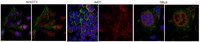
Immunocytochemistry Analysis: 1:500 dilution from a representative lot detected NET29 in NIH/3T3, A431, and HeLa cells.
Immunofluorenscence Analysis: A representative lot was used by an independent laboratory in IF. (Malik, P., et al. (2010).
Biological Information
Immunogen
KLH-conjugated linear peptide corresponding to human NET29 at the extracellular domain.
Epitope
Extracellular domain
Concentration
Please refer to the Certificate of Analysis for the lot-specific concentration.
Host
Rabbit
Specificity
This antibody recognizes NET29 at the extracellular domain.
SUBCELLULAR LOCATION: Membrane; Multi-pass membrane protein Potential.
SEQUENCE SIMILARITIES: Belongs to the TMEM120 family.
Molecular Weight
Two different isoforms observed at ~ 41 kDa (isoform 1) and ~35 kDa (isoform 2).
Physicochemical Information
Dimensions
Materials Information
Toxicological Information
Safety Information according to GHS
Safety Information
Product Usage Statements
Quality Assurance
Evaluated by Western Blot in rat liver tissue lysate.
Western Blot Analysis: 0.5 µg/mL of this antibody detected NET29 on 10 µg of rat liver tissue lysate.
Usage Statement
Unless otherwise stated in our catalog or other company documentation accompanying the product(s), our products are intended for research use only and are not to be used for any other purpose, which includes but is not limited to, unauthorized commercial uses, in vitro diagnostic uses, ex vivo or in vivo therapeutic uses or any type of consumption or application to humans or animals.
TMEM120A and B: Nuclear Envelope Transmembrane Proteins Important for Adipocyte Differentiation. Batrakou, DG; de Las Heras, JI; Czapiewski, R; Mouras, R; Schirmer, EC PloS one
10
e0127712
2015
Recent work indicates that the nuclear envelope is a major signaling node for the cell that can influence tissue differentiation processes. Here we present two nuclear envelope trans-membrane proteins TMEM120A and TMEM120B that are paralogs encoded by the Tmem120A and Tmem120B genes. The TMEM120 proteins are expressed preferentially in fat and both are induced during 3T3-L1 adipocyte differentiation. Knockdown of one or the other protein altered expression of several genes required for adipocyte differentiation, Gata3, Fasn, Glut4, while knockdown of both together additionally affected Pparg and Adipoq. The double knockdown also increased the strength of effects, reducing for example Glut4 levels by 95% compared to control 3T3-L1 cells upon pharmacologically induced differentiation. Accordingly, TMEM120A and B knockdown individually and together impacted on adipocyte differentiation/metabolism as measured by lipid accumulation through binding of Oil Red O and coherent anti-Stokes Raman scattering microscopy (CARS). The nuclear envelope is linked to several lipodystrophies through mutations in lamin A; however, lamin A is widely expressed. Thus it is possible that the TMEM120A and B fat-specific nuclear envelope transmembrane proteins may play a contributory role in the tissue-specific pathology of this disorder or in the wider problem of obesity.
Specific nuclear envelope transmembrane proteins can promote the location of chromosomes to and from the nuclear periphery. Zuleger, N; Boyle, S; Kelly, DA; de las Heras, JI; Lazou, V; Korfali, N; Batrakou, DG; Randles, KN; Morris, GE; Harrison, DJ; Bickmore, WA; Schirmer, EC Genome biology
14
R14
2013
Different cell types have distinctive patterns of chromosome positioning in the nucleus. Although ectopic affinity-tethering of specific loci can be used to relocate chromosomes to the nuclear periphery, endogenous nuclear envelope proteins that control such a mechanism in mammalian cells have yet to be widely identified.To search for such proteins, 23 nuclear envelope transmembrane proteins were screened for their ability to promote peripheral localization of human chromosomes in HT1080 fibroblasts. Five of these proteins had strong effects on chromosome 5, but individual proteins affected different subsets of chromosomes. The repositioning effects were reversible and the proteins with effects all exhibited highly tissue-restricted patterns of expression. Depletion of two nuclear envelope transmembrane proteins that were preferentially expressed in liver each reduced the normal peripheral positioning of chromosome 5 in liver cells.The discovery of nuclear envelope transmembrane proteins that can modulate chromosome position and have restricted patterns of expression may enable dissection of the functional relevance of tissue-specific patterns of radial chromosome positioning.