Wenn Sie das Fenster schließen, wird Ihre Konfiguration nicht gespeichert, es sei denn, Sie haben Ihren Artikel in die Bestellung aufgenommen oder zu Ihren Favoriten hinzugefügt.
Klicken Sie auf OK, um das MILLIPLEX® MAP-Tool zu schließen oder auf Abbrechen, um zu Ihrer Auswahl zurückzukehren.
Wählen Sie konfigurierbare Panels & Premixed-Kits - ODER - Kits für die zelluläre Signaltransduktion & MAPmates™
Konfigurieren Sie Ihre MILLIPLEX® MAP-Kits und lassen sich den Preis anzeigen.
Konfigurierbare Panels & Premixed-Kits
Unser breites Angebot enthält Multiplex-Panels, für die Sie die Analyten auswählen können, die am besten für Ihre Anwendung geeignet sind. Unter einem separaten Register können Sie das Premixed-Cytokin-Format oder ein Singleplex-Kit wählen.
Kits für die zelluläre Signaltransduktion & MAPmates™
Wählen Sie gebrauchsfertige Kits zur Erforschung gesamter Signalwege oder Prozesse. Oder konfigurieren Sie Ihre eigenen Kits mit Singleplex MAPmates™.
Die folgenden MAPmates™ sollten nicht zusammen analysiert werden: -MAPmates™, die einen unterschiedlichen Assaypuffer erfordern. -Phosphospezifische und MAPmate™ Gesamtkombinationen wie Gesamt-GSK3β und Gesamt-GSK3β (Ser 9). -PanTyr und locusspezifische MAPmates™, z.B. Phospho-EGF-Rezeptor und Phospho-STAT1 (Tyr701). -Mehr als 1 Phospho-MAPmate™ für ein einziges Target (Akt, STAT3). -GAPDH und β-Tubulin können nicht mit Kits oder MAPmates™, die panTyr enthalten, analysiert werden.
.
Bestellnummer
Bestellinformationen
St./Pkg.
Liste
Dieser Artikel wurde zu Ihren Favoriten hinzugefügt.
Wählen Sie bitte Spezies, Panelart, Kit oder Probenart
Um Ihr MILLIPLEX® MAP-Kit zu konfigurieren, wählen Sie zunächst eine Spezies, eine Panelart und/oder ein Kit.
Custom Premix Selecting "Custom Premix" option means that all of the beads you have chosen will be premixed in manufacturing before the kit is sent to you.
Catalogue Number
Ordering Description
Qty/Pack
List
Dieser Artikel wurde zu Ihren Favoriten hinzugefügt.
Spezies
Panelart
Gewähltes Kit
Menge
Bestellnummer
Bestellinformationen
St./Pkg.
Listenpreis
96-Well Plate
Menge
Bestellnummer
Bestellinformationen
St./Pkg.
Listenpreis
Weitere Reagenzien hinzufügen (MAPmates erfordern die Verwendung eines Puffer- und Detektionskits)
Menge
Bestellnummer
Bestellinformationen
St./Pkg.
Listenpreis
48-602MAG
Buffer Detection Kit for Magnetic Beads
1 Kit
Platzsparende Option Kunden, die mehrere Kits kaufen, können ihre Multiplex-Assaykomponenten in Kunststoffbeuteln anstelle von Packungen erhalten, um eine kompaktere Lagerung zu ermöglichen.
Dieser Artikel wurde zu Ihren Favoriten hinzugefügt.
Das Produkt wurde in Ihre Bestellung aufgenommen
Sie können nun ein weiteres Kit konfigurieren, ein Premixed-Kit wählen, zur Kasse gehen oder das Bestell-Tool schließen.
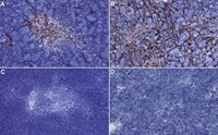
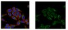
Anti-Medullary epithelium, clone ER-TR5, Cat. No. MABF2818, is a rat monoclonal antibody that detects Medullary epithelium and is tested for use in Immunohistochemistry and Immunofluorescence.
More>>Anti-Medullary epithelium, clone ER-TR5, Cat. No. MABF2818, is a rat monoclonal antibody that detects Medullary epithelium and is tested for use in Immunohistochemistry and Immunofluorescence. Less<<
The thymus is considered as the primary lymphoid organ that is responsible for the generation and maturation of T cells. Thymic epithelial cells account for the majority of thymic stromal components. These cells can be divided into cortical and medullary thymic epithelial cells based on their localization within the thymus. Cortical thymic epithelial cells are essential for the positive selection of T cells, whereas the medullary thymic epithelial cells (mTEC) play a role in inducing negative selection of highly reactive T cells that are required for establishing central self-tolerance. They participate in self-tolerance by eliminating self-reactive T cells. In murine species, mTEC are further divided into mTEC low and mTEC high according to the expression levels of several maturation molecules, such as MHCII and CD8o. mTEC low cells can serve as precursors for mature mTEC high cells. mTECs have also been classified into four major subsets, termed mTEC I-IV depending on their distinct transcriptional and molecular characteristics. mTEC I and II subpopulation appear early and can be detected at E18.5 and are most proliferative cells. mTEC III are detected in thymus at 4-weeks. mTEC IV are visible on day 6 in neonatal mice. The function of mTECs is dependent on their characteristic features such as ectopic expression of peripheral tissue restricted antigens and their master regulator-autoimmune regulator (Aire), expression of various chemokines and cytokines. Lymphotoxin β receptor (LTβR) is reported to be an essential regulator of multiple mTEC subsets within the mTEC low compartment. It controls thymic tolerance by regulating the frequency and makeup of intrathymic dendritic cells that are required for effective thymocyte negative selection. Clone ER-TR5 is shown to exclusively react with mTECs. (Ref.: Wang, HX., et al. (2020). Front. Immunol. 10; 3099; Lucas, B., et al. (2020). Nat. Commun. 11; Article 2198; Cosway, EJ., et al. (2017). J. Exp. Med. 214(11); 3183-3195; Van Vliet, E., et al. (1984). Eur. J. Immunol. 14(6); 524-529).
References
Product Information
Format
Purified
Presentation
Purified rat monoclonal antibody IgM in buffer containing 0.1 M Tris-Glycine (pH 7.4), 150 mM NaCl with 0.05% sodium azide.
Anti-Medullary epithelium, clone ER-TR5, Cat. No. MABF2818, is a rat monoclonal antibody that detects Medullary epithelium and is tested for use in Immunohistochemistry and Immunofluorescence.
Key Applications
Immunohistochemistry
Immunofluorescence
Application Notes
Tested applications
Immunofluorescence Analysis: A representative lot detected Medullary epithelium in Immunofluorescence applications (Akiyama, T., et al. (2005). Science. 308(5719):248-51; Lei, Y., et al. (2011). J Exp Med. 208(2):383-94; Cosway, E.J., et al. (2017). J Exp Med. 214(11):3183-3195).
Immunohistochemistry Applications: A representative lot detected Medullary epithelium in Immunohistochemistry applications (Van Vliet, E., et al. (1984). Eur J Immunol. 14(6):524-9; Akiyama, T., et al. (2005). Science.;308(5719):248-51; Dooley, J., et al. (2006). J Immunol. 176(11):6484-90; Lei, Y., et al. (2011). J Exp Med. 208(2):383-94; Cosway, E.J., et al. (2017). J Exp Med. 214(11):3183-3195).
Note: Actual optimal working dilutions must be determined by end user as specimens, and experimental conditions may vary with the end user
Biological Information
Immunogen
Thymic stromal cells isolated from C3H mice.
Epitope
Unknown
Clone
ER-TR5
Concentration
1.0 mg/mL. Please refer to guidance on suggested starting dilutions and/or titers per application and sample type.
Host
Rat
Specificity
Clone ER-TR5 is a rat monoclonal antibody that detects Medullary epithelial cells.
Evaluated by Immunohistochemistry in Mouse thymus tissue.
Immunohistochemistry Applications: 1:150 dilution of this antibody detected Medullary epithelium in Mouse thymus tissue sections.
Usage Statement
Unless otherwise stated in our catalog or other company documentation accompanying the product(s), our products are intended for research use only and are not to be used for any other purpose, which includes but is not limited to, unauthorized commercial uses, in vitro diagnostic uses, ex vivo or in vivo therapeutic uses or any type of consumption or application to humans or animals.