Wenn Sie das Fenster schließen, wird Ihre Konfiguration nicht gespeichert, es sei denn, Sie haben Ihren Artikel in die Bestellung aufgenommen oder zu Ihren Favoriten hinzugefügt.
Klicken Sie auf OK, um das MILLIPLEX® MAP-Tool zu schließen oder auf Abbrechen, um zu Ihrer Auswahl zurückzukehren.
Wählen Sie konfigurierbare Panels & Premixed-Kits - ODER - Kits für die zelluläre Signaltransduktion & MAPmates™
Konfigurieren Sie Ihre MILLIPLEX® MAP-Kits und lassen sich den Preis anzeigen.
Konfigurierbare Panels & Premixed-Kits
Unser breites Angebot enthält Multiplex-Panels, für die Sie die Analyten auswählen können, die am besten für Ihre Anwendung geeignet sind. Unter einem separaten Register können Sie das Premixed-Cytokin-Format oder ein Singleplex-Kit wählen.
Kits für die zelluläre Signaltransduktion & MAPmates™
Wählen Sie gebrauchsfertige Kits zur Erforschung gesamter Signalwege oder Prozesse. Oder konfigurieren Sie Ihre eigenen Kits mit Singleplex MAPmates™.
Die folgenden MAPmates™ sollten nicht zusammen analysiert werden: -MAPmates™, die einen unterschiedlichen Assaypuffer erfordern. -Phosphospezifische und MAPmate™ Gesamtkombinationen wie Gesamt-GSK3β und Gesamt-GSK3β (Ser 9). -PanTyr und locusspezifische MAPmates™, z.B. Phospho-EGF-Rezeptor und Phospho-STAT1 (Tyr701). -Mehr als 1 Phospho-MAPmate™ für ein einziges Target (Akt, STAT3). -GAPDH und β-Tubulin können nicht mit Kits oder MAPmates™, die panTyr enthalten, analysiert werden.
.
Bestellnummer
Bestellinformationen
St./Pkg.
Liste
Dieser Artikel wurde zu Ihren Favoriten hinzugefügt.
Wählen Sie bitte Spezies, Panelart, Kit oder Probenart
Um Ihr MILLIPLEX® MAP-Kit zu konfigurieren, wählen Sie zunächst eine Spezies, eine Panelart und/oder ein Kit.
Custom Premix Selecting "Custom Premix" option means that all of the beads you have chosen will be premixed in manufacturing before the kit is sent to you.
Catalogue Number
Ordering Description
Qty/Pack
List
Dieser Artikel wurde zu Ihren Favoriten hinzugefügt.
Spezies
Panelart
Gewähltes Kit
Menge
Bestellnummer
Bestellinformationen
St./Pkg.
Listenpreis
96-Well Plate
Menge
Bestellnummer
Bestellinformationen
St./Pkg.
Listenpreis
Weitere Reagenzien hinzufügen (MAPmates erfordern die Verwendung eines Puffer- und Detektionskits)
Menge
Bestellnummer
Bestellinformationen
St./Pkg.
Listenpreis
48-602MAG
Buffer Detection Kit for Magnetic Beads
1 Kit
Platzsparende Option Kunden, die mehrere Kits kaufen, können ihre Multiplex-Assaykomponenten in Kunststoffbeuteln anstelle von Packungen erhalten, um eine kompaktere Lagerung zu ermöglichen.
Dieser Artikel wurde zu Ihren Favoriten hinzugefügt.
Das Produkt wurde in Ihre Bestellung aufgenommen
Sie können nun ein weiteres Kit konfigurieren, ein Premixed-Kit wählen, zur Kasse gehen oder das Bestell-Tool schließen.
Anti-MART-1/Melan-A, clone A103, Cat. No. MABF3201, is a mouse monoclonal antibody that detects MART-1 and is tested for use in Immunohistochemistry and Western Blotting.
More>>Anti-MART-1/Melan-A, clone A103, Cat. No. MABF3201, is a mouse monoclonal antibody that detects MART-1 and is tested for use in Immunohistochemistry and Western Blotting. Less<<
Empfohlene Produkte
Übersicht
Replacement Information
Description
Catalogue Number
MABF3201-100UL
Description
Anti-MART-1/Melan-A Antibody, clone A103
Alternate Names
Melanoma antigen recognized by T-cells 1
Antigen LB39-AA
Antigen SK29-AA
Protein Melan-A
Background Information
Melanoma antigen recognized by T-cells 1 (UniProt: Q16655; also known as MART-1, Antigen LB39-AA, Antigen SK29-AA, Protein Melan-A) is encoded by the MLANA (also known as MART1) gene (Gene ID: 2315) in human. MART-1, a melanosome-specific protein, is a single-pass type III membrane protein with a transmembrane domain (aa 27-47) and a cytoplasmic domain (aa 48-118). Its expression is restricted to melanocytes, melanoma, and retina and is absent in other tissues and tumors. It is highly enriched in early melanosomes (Stage I and/or II melanosomes) indicating that its role in early melanogenesis. It is reported to stabilize both GPCR143/OA-1 and plays a role in the expression, stability, trafficking, and processing of pre-melanosome protein (PMEL) to ensure the formation of a stage 2 melanosome. It is mainly found in the endoplasmic reticulum (ER) and Golgi but is also detected in small vesicles and tubules dispersed over the entire cytoplasm. A small fraction of the protein is inserted into the membrane in an inverted orientation, which allows melanoma cells to be effectively recognized by specific cytotoxic T-cells. MART-1 may be a useful marker in the diagnosis of melanocytic lesions and in the differential diagnosis of malignant melanoma. (Ref.: Hoashi, T., et al. (2005). J. Biol. Chem. 280(14); 14006-14016; Chen, YT., et al. (1996). Proc. Natl. Acad. Sci. USA. 93(12); 5915-5919).
References
Product Information
Format
Purified
Presentation
Purified mouse monoclonal antibody IgG1 in buffer containing 0.1 M Tris-Glycine (pH 7.4), 150 mM NaCl with 0.05% sodium azide.
Anti-MART-1/Melan-A, clone A103, Cat. No. MABF3201, is a mouse monoclonal antibody that detects MART-1 and is tested for use in Immunohistochemistry and Western Blotting.
Key Applications
Western Blotting
Immunohistochemistry
Application Notes
Tested applications
Western Blotting Analysis: A representative lot detected MART-1 in Western Blotting applicaiton. (Chen, Y.T., et al. (1996). Proc Natl Acad Sci USA. 93(12); 5915-9).
Immunohistochemistry Applications: A representative lot detected MART-1/Melan-A in Immunohistochemistry applications (Chen, Y.T., et al. (1996). Proc Natl Acad Sci USA. 93(12);5915-9).
Note: Actual optimal working dilutions must be determined by end user as specimens, and experimental conditions may vary with the end user.
Biological Information
Immunogen
Full-length, recombinant, human MART-1 produced in E.coli.
Epitope
Unknown
Clone
A103
Concentration
0.5 mg/mL. Please refer to guidance on suggested starting dilutions and/or titers per application and sample type.
Host
Mouse
Specificity
Clone A103 is a mouse monoclonal antibody that detects MART-1.
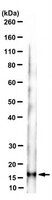
~16 kDa observed; 13.16 kDa calculated. Uncharacterized bands may be observed in some lysate(s).
Physicochemical Information
Dimensions
Materials Information
Toxicological Information
Safety Information according to GHS
Safety Information
Product Usage Statements
Quality Assurance
Evaluated by Western Blotting in SK-MEL-28 cell lysate.
Western Blotting Analysis: A 1:500 dilution of this antibody detected MART-1 in SK-MEL-28 cell lysate.
Usage Statement
Unless otherwise stated in our catalog or other company documentation accompanying the product(s), our products are intended for research use only and are not to be used for any other purpose, which includes but is not limited to, unauthorized commercial uses, in vitro diagnostic uses, ex vivo or in vivo therapeutic uses or any type of consumption or application to humans or animals.